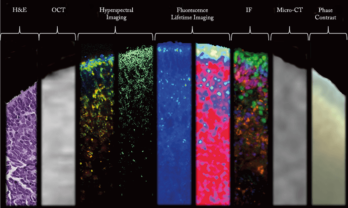
Andrew Browne, a former resident at the University of Southern California Roski Eye Institute, USA, sent us this composite image of a mature 3D retinal organoid. In their study, the team used a range of non-invasive imaging techniques to study the growth and development of the retinal organoids, which were derived from human pluripotent stem cells (hPSCs). “This composite highlights the seven different imaging modalities used in the study, in which fluorescence lifetime imaging and hyperspectral imaging provided new insights into, and techniques to understand, organoid biology,” says Browne. Credit: AW Browne et al., Invest Ophthalmol Vis Sci, 58, 3311–3318 (2017).
