You might not think that one of the most challenging aspects of eye imaging is obtaining a clear picture of the blood vessels in the retina. After all, the retina is thin and nearly transparent, and yet – despite over half a century of retinal imaging – our most commonly used method for doing so still has major limitations.
Fluorescein angiography (FA) is today’s standard imaging method for examining retinal blood circulation. With it, ophthalmologists can easily see superficial blood vessels, but two out of the three major capillary networks in the retina remain largely a mystery; both the radial peripapillary capillaries and the inner capillary network are poorly visualized with this method. The retinal capillaries that are seen in fluorescein angiograms are set against a backdrop of low-level fluorescence that’s often ascribed to the choroid, but may actually originate from the deeper capillary plexus. Why can’t these deeper layers of the retinal vasculature be seen clearly? The most likely reason for this is that retinal nerve fiber bundles scatter light, leading to a diffuse glow instead of a sharply defined fluorescent image.
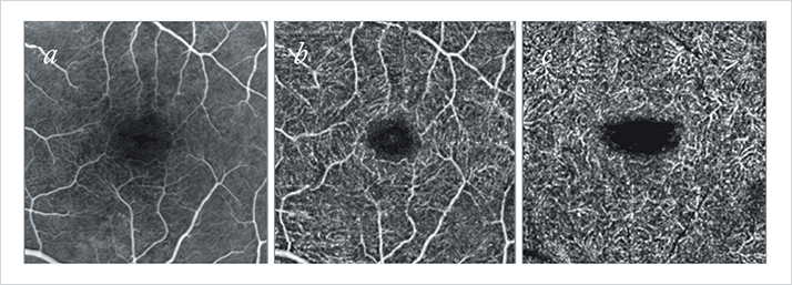
OCT angiography is a relatively new alternative to FA; it requires little in the way of patient preparation and involves no dye injections – avoiding the rare, but real risk of dye-related adverse events. The OCT part is the rapid scanning of regions of retinal tissue; the angiography part is the algorithmic analysis of those images, identifying variations between scans in measures of reflectivity, phase shift, or phase variance – which enables the construction of microvascular flow maps. Three ophthalmologists from Vitreous-Retina-Macula Consultants of New York compared OCT angiography with FA imaging in the eyes of 12 study participants (1). Not surprisingly, they found that FA failed to image the radial peripapillary or deep capillary networks in any of the twelve eyes. OCT angiography, however, successfully imaged all layers of the retinal vasculature in all of the study participants’ eyes (Figure 1).
OCT angiography isn’t likely to replace FA imaging in the short term – FA is a well-known, well-characterized method, and OCT angiography has a number of limitations beyond its relative infancy in terms of characterization (Table 1). But the technique will evolve, improve and should be increasingly adopted. The study authors suggest that OCT angiography will help provide better characterizations of the vascular changes that occur over time in many common retinal diseases.
References
- R.F. Spaide, J.M. Klancnik Jr, M.J. Cooney, “Retinal vascular layers imaged by fluorescein angiography and optical coherence tomography angiography”, JAMA Ophthalmol. Epub ahead of print (2014). doi: 10.1001/jamaophthalmol.2014.3616.
