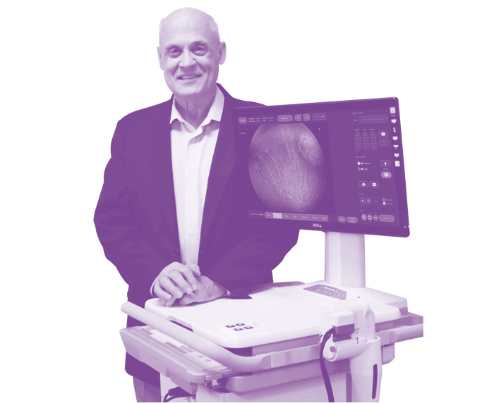
Bert Massie worked in the aerospace industry for many years, developing his knowledge and expertise in adaptive optics and interferometry. At the height of his career, he followed a calling: advancing the field of ophthalmology by responding to the most pressing needs for retinal imaging. Since then, he has developed the RetCam, used worldwide in screening for retinopathy of prematurity, the Micron for eye research, and, more recently, the ICON – an improved wide-angle imaging system. Here, he shares his passion for entrepreneurship, and describes how innovations in ophthalmology have been driven by the evolution of sensors, microprocessors and lasers.
A curious mind
I’m a scientist and an engineer but I tend to think of myself as an entrepreneur. I don’t think that this is something you learn – it is a natural and intense personal drive. It is evident in everything I do; it gets me out of bed early in the morning.
As a young child I was very curious about how things worked. In early grade school I took my toy electric train engine apart because I really wanted to know what all those gears did. I developed an interest in astronomy, and for many nights lay outside on a blanket to watch the stars appear in the evening sky. I knew all the constellations. For an entrepreneur curiosity is foundational, but must be complemented by a creative mind, and a self-confidence and drive to develop the ideas that originate in your mind. For example, as a teenager I built a 12” telescope to explore an idea about how to obtain color images of galaxies.
Aerospace adventures
My first real “creative” step was my PhD thesis, where I tested new ideas to measure the non-linear properties of solids using ultrasound with laser probes. Post graduate school I entered the aerospace field, and for 11 years I was involved in the Strategic Defense Initiative, nicknamed Star Wars, and in my last aerospace role I spent five years working in a senior position at the Lawrence Livermore National Laboratory, which is a federal research facility in California, US, focusing on finding solutions to security-related challenges. In my aerospace career I was awarded over 25 patents in optics, I published the same number of journal articles, and I edited a reference book on optical technology. I developed techniques for interferometry – classically, interferometers make accurate measurements of optical components – and my most interesting project was a high-performance two-wavelength interferometer. Optical Coherence Tomography uses multiple wavelengths, but at that time the required “low-coherence” sources did not exist, so I had to use two lasers, which required a high level of sophistication. Following this, I developed a novel adaptive mirror for correction of atmospheric aberrations on imaging of space objects.
In my last position with the national laboratory, together with my colleagues, I developed a technique to image space optics through the turbulent atmosphere without using adaptive optics. This technique was proven in experiments between an object on a mountain top and a lower altitude telescope.
I enjoyed working in the aerospace industry, but from the very beginning of my career – since my undergraduate years, in fact – I sought an opportunity where I could help people with vision impairment regain their sight. I sought a role where I could focus on more meaningful goals.
Problems and solutions
When I decided to make a career in ophthalmology, I found that much of my extensive optics knowledge and experience was applicable. I started working on various ophthalmic solutions, including a non-contact ultrasound-based tonometer, which worked, but was too expensive to merchandise. As it often happens, through a random series of events, I met a very prominent physician - A. Linn Murphree - who introduced me to the challenge of wide-field imaging in children. The device developed by Dr. Murphree and his team did not adequately perform, and I picked where they left off. I redesigned the instrument from the ground up, and this became the RetCam.
RetCam was originally developed to image retinoblastoma but was quickly adapted to image retinopathy of prematurity (ROP). This disorder potentially afflicts infants born before 31 weeks of gestation and weighing less than 1250 g. There is significant ocular morbidity associated with ROP, which usually develops in both eyes. It is among the most common causes of childhood vision loss and often leads to permanent vision impairment.
We were told that we would sell a dozen units, but the RetCam was widely adopted and there are now over 2,000 RetCam units in use around the world. It can be operated by non-ophthalmologists, who can send images to a central facility, where they are professionally evaluated. It greatly reduces the labor costs and the time it takes for the problematic cases to be picked up. A point that is especially vital in developing countries; there are very competent clinicians working there, but they don’t always have the staff to screen every child. If diagnosis and treatment is timely, it is usually very effective and protects patients from lifelong blindness.
Answering the call
When I left the RetCam program, I focused on developing technology for eye and eye-brain research, using laboratory animals such as mice and rats, and introduced a retinal imaging microscope called the Phoenix Micron. Developing the mouse imaging system was quite difficult and there were times where I was certain that failure was just a day away. Nevertheless, the Phoenix Micron retinal imaging microscope became a reality. I was again told that I might be able to sell a dozen units – and we now have over 300 units in use in major institutions around the world.
At the age of 71, when I was about to retire, and with pressure from several sources, including several senior clinical leaders – I decided to return to clinical ophthalmology and develop a successor to the RetCam. The base technology for the RetCam had not changed since inception. Ophthalmologists had been complaining about issues with imaging patients from ethnic minority groups – those with darkly pigmented retinas – such as African Americans and Asians. The RetCam performed poorly with dark retinas and could not image adults. When the retina is dark, the light returned to the image sensor is lower than the scatter in the cornea. As a result, the image is lost in the haze.
In developing what became the Phoenix ICON my team and I accepted an absolute directive to develop high-contrast/high-resolution imaging even in darkly pigmented retinas, and in the process were able to also image adults – something RetCam had not been able to do. The Phoenix ICON design was a complete change from the RetCam and arose from a “blank sheet of white paper.” Getting the light in and to the retina without the scatter is quite difficult – these difficulties were overcome, but only by ignoring a number of classic ophthalmic rules along the way!
The Phoenix ICON lived up to its objectives and is a wide-angle retinal imaging system with high-contrast imaging and ability to image adults as well. I believe Phoenix ICON’s capability has the potential to contribute to new areas such as melanoma; all this is the subject of clinical trials for validation. We also have plans to provide the technology to countries requiring humanitarian assistance: we’ve been talking to The Queens Jubilee Trust in the UK, who can help us answer the needs for these systems in India.
Technological evolution of the last few decades has really influenced ophthalmology. When sensors, such as CCD or CMOS, entered the commercial market, the performance of instrumentation improved considerably. The sensor in the Phoenix ICON allows the provision of much lower light level onto the retina, enabling imaging of awake adult patients.
More powerful microprocessors have also had a major impact, and at first there wasn’t a good way of handling digital data. Storage has also seen unbelievable gains in volume and affordability. The first RetCam had 600 MB of optical storage; now, we can store thousands of images. And we can transfer large data files easily and store images in the cloud – something that we had not considered previously.
Lasers are another example of seismic change. As a graduate student we built our own lasers, and now they are readily available to applications such as ophthalmology.
Working backwards
I begin my projects by first being certain that the right question is identified. For example, many times RetCam users asked for more resolution, but they really needed improved visibility, meaning higher contrast. Innovations such as the Phoenix ICON and Phoenix Micron need to not only be a technological advancement, but also an advancement that makes a useful difference. Accordingly, as a final and acid test before I launch a project, I imagine myself standing in front of the physician and showing the device; if I do not see excitement from the user, I do not proceed.
Innovations must make a difference, not just be different, and that is one of the most important reasons for the decision on pursuing a project. This means of evaluation has served me well.
N.A.(Bert) Massie, Ph.D., has more than 40 years of experience as a lead entrepreneur of a variety of optical-based technologies. He is currently working for the Phoenix Technology Group.
