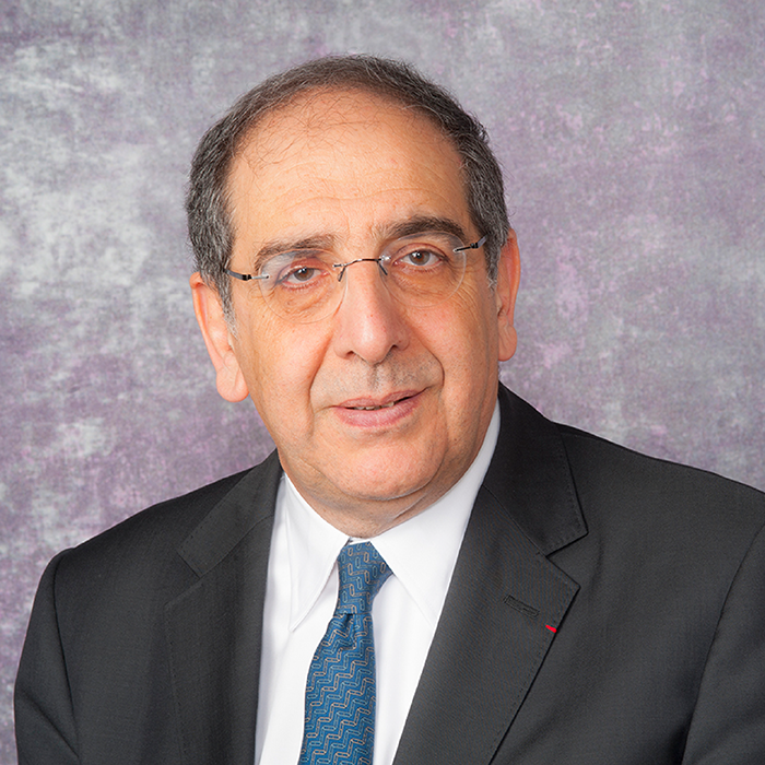
The International Prize for Translational Neuroscience has honored and promoted outstanding biomedical scientists and clinicians since its introduction in 1990. With close ties to the Max Planck Society, the award is endowed with a €60,000 prize. The most recent award recognizes two pioneering researchers: José-Alain Sahel, Chair of the Department of Ophthalmology at the University of Pittsburgh School of Medicine, and Botond Roska, Founding Director and Senior Group Leader at the Institute for Molecular and Clinical Ophthalmology Basel.
The pair have collaborated on research projects for over 20 years, but it was their optogenetic breakthrough that caught the attention of the committee. Specifically, they used optogenetic methods to partially restore vision in patients blinded by retinitis pigmentosa (RP), representing the first example of optogenetics in humans and a milestone in the treatment of blinding conditions that affect millions of people worldwide.
We spoke with José-Alain Sahel to discuss what this award means for him and his future in ophthalmology.
What does winning this award mean for you?
The list of previous awardees is very impressive and, as an ophthalmologist, I was thrilled to join this outstanding group of scientists. Translation is at the core of my passion. Ophthalmologists should feel proud that ophthalmology is the first field where optogenetics has demonstrated clinically relevant results.
The award also means recognizing my collaboration with Botond Roska – my close friend and an exceptional scientist with a breadth of expertise spanning neuroscience, logical thinking, and medicine – as well as the years of work from my Paris and Pittsburgh teams. Although the award is only delivered to a few, there are many people who contributed to this achievement.
Please explain the underlying principles of optogenetics…
Optogenetics is based on the expression of proteins, identified in algae a couple of decades ago, able to respond to light activation by eliciting a transmembrane electrical current. By expressing such proteins, any cell can become light sensitive. These proteins are robust and can be used to study any neuronal circuitry. This tool has transformed neuroscience.
How was optogenetics applied in your research to restore vision in blind patients?
Because the expression of these types of proteins (by gene therapy) can transform any cell in the retina into a photoreceptor, a few of us envisioned that this could be used to restore visual responses in degenerated retinas affected by various genetic defects – leading to the loss of photoreceptor function.
What specific challenges does the application of optogenetics to ophthalmology present?
These proteins are robust, but require a strong level of light stimulation. Natural phototransduction is able to respond to any level of lighting and we need to restore both light sensitivity and the ability to respond to a wide range of light intensities, as well as emitting patterns that can be interpreted meaningfully. A device emitting light in the relevant wavelength is usually required to provide enough stimulation, but also to compensate for the lack of adaptation of these proteins. The device (goggles mounted with a camera and light projecting system) is currently bulky, but it provides controllable stimulation.
Some groups and companies are using proteins that may not require the device for stimulation but the issues of pattern stimulation and adaptation to the wide range of lighting conditions remain to be addressed.
Given that a high level of illumination is required the risk of light toxicity must be addressed. To that effect, we have identified, tested, and validated red-shifted proteins that provide a very good safety profile. ChrimsonR was engineered by Ed Boyden at MIT. These proteins are totally foreign to the human immune system and so the risk of inflammation and immune response, already significant in gene therapy, must be considered.
Finally, most strategies stimulate both the ON and the OFF responses, which reduces the resolution and affects image processing. The trial we conducted addressed several of these concerns and provided very promising initial data.
What are you working on now?
Due to the unique nature of our findings, we wanted to verify the validity using several methods: multielectrode electro-encephalography to demonstrate cortical activation, paired with reporting by the patient. The trial is still ongoing with an extension cohort as the dose escalation established the safety.
Currently, we are working on cell-specific targeting, improved delivery and specific encoding to achieve a better resolution. We are also exploring other cell types (dormant cone photoreceptors, bipolar cells) and possibly a combination with cell therapy.
This work is conducted within our academic labs and in partnership with companies, mainly GenSight (ganglion cell stimulation), but also Sparing Vision (cone cell stimulation) and Tenpoint – a company that combines optogenetics and cell therapy, which I cofounded.
What other research projects have given you a sense of pride?
My focus is on patient centered care; I run clinics every week dedicated to these patients. This keeps me from feeling too proud as we still have a way to go before we achieve our goals. Patient expectations help fuel our work on several approaches to fighting blindness in retinal degenerations. To list a few projects: I have been fortunate enough to work on corrective gene therapies, neuroprotection of cone photoreceptors, optogenetics, cell therapy, and I have been at the forefront of the development of the PRIMA wireless subretinal photovoltaic implant.
I am mostly proud of being persistent in leading these projects over decades, despite numerous challenges. Even more importantly, I feel privileged to work with very talented and dedicated colleagues from both academia and industry. Building teams has been a key driver, alongside building research institutes from scratch, such as the Institut de la Vision in Paris and the UPMC Vision Institute in Pittsburgh.
What are the next steps for your career?
After founding and directing the Institut de la Vision in Paris as well as chairing two Departments of Ophthalmology (National Ophthalmology Hospital-/Quinze-Vingts; Fondation Ophtalmologique Rothschild), I am now chairing the Department of Ophthalmology at the University of Pittsburgh and directing the brand new 400,000 square foot Vision Institute built by UPMC.
We just opened this superb ten-story building that integrates clinical care, comprehensive and all subspecialties, education (including a large surgical training laboratory), translational and basic research, and technology transfer. Developing new therapies, testing innovations including AI, improving access to care for all are top priorities for the faculty (80+) and staff here.
My own research is continuing along the axes I described above, but I am particularly interested in understanding and characterizing the impact of visual impairment and vision restoration on daily life and patient experience – using patient-reported outcomes, performance-based tests, narrative research, including phenomenology and art.
If you weren’t involved in neuroscience, what would you be doing?
I am primarily an ophthalmologist and only an amateur neuroscientist! If I weren’t involved in these, I would be playing music and writing – as an amateur also! And I would have more time to dedicate to my family and friends.
