Conexiant
Login
  • Corneal Physician
  • Glaucoma Physician
  • New Retinal Physician
  • Ophthalmology Management
  • Ophthalmic Professional
  • Presbyopia Physician
  • Retinal Physician
The Ophthalmologist
  • Explore

    Explore

    • Latest
    • Insights
    • Case Studies
    • Opinion & Personal Narratives
    • Research & Innovations
    • Product Profiles

    Featured Topics

    • Anterior Segment
    • Glaucoma
    • Retina

    Issues

    • Latest Issue
    • Archive
  • Subspecialties
    • Cataract
    • Cornea
    • Glaucoma
    • Neuro-ophthalmology
    • Oculoplastics
    • Pediatric
    • Retina
  • Business

    Business & Profession

    • Professional Development
    • Business and Entrepreneurship
    • Practice Management
    • Health Economics & Policy
  • Training & Education

    Career Development

    • Professional Development
    • Career Pathways

    Events

    • Webinars
    • Live Events
  • Events
    • Live Events
    • Webinars
  • Community

    People & Profiles

    • Power List
    • Voices in the Community
    • Authors & Contributors
  • Multimedia
    • Video
    • Podcasts
Subscribe
Subscribe

False

Advertisement
The Ophthalmologist / Issues / 2014 / Sep / Feeling the Pressure
Anterior Segment Cataract Glaucoma

Feeling the Pressure

Can a new implantable microfluidic sensor put IOP tracking in the hands – or rather eyes – of patients with glaucoma?

By Roisin McGuigan 9/9/2014 1 min read

Share

Zeiss-SMILE-Skyscraper-160x600
Zeiss-SMILE-Leaderboard-728x90
Zeiss-SMILE-Skyscraper-160x600

American and Israeli researchers have developed an implantable sensor that they say measures intraocular pressure (IOP) with high accuracy and reproducibility – when tested in a porcine model (1). IOP is notorious for fluctuating over the course of the day, which makes getting a truly representative measurement with tonometry tricky; posture makes a difference and peak IOP is often not at the time of measurement – it’s when the patient is lying on their back sleeping at night. Furthermore, Goldmann applanation tonometry requires topical anesthesia and can be affected by variations in corneal biomechanical properties, such as thickness or disease state.

0814-205-upfront-mainFigure 1. The IOP-reading IOL. a. The air/fluid interface can be easily captured by a smartphone with an optical adaptor; b. Scales can be incorporated onto the outer side of the channel to help measure the location of the air/fluid interface; c. The sensor can be embedded in an IOL and implanted during cataract surgery. Image credit: Ismail Araci, Baolong Su, Stephen Quake and Yossi Mandel.

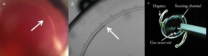
The new device uses a passive pressure sensor implant that relies on microfluidic physics principles. In other words, the implant contains a tiny open channel that draws in the aqueous intraocular fluid (due to capillary forces and IOP), that results in the compression of a gas reservoir attached to the channel. The greater the IOP, the more compressed the gas becomes. To calculate IOP, the relative position of the aqueous-gas interface can be measured with a specialized camera, a slit lap during a routine eye examination – or even a smartphone equipped with an adaptor. The device was originally developed as a stand-alone implant (inserted through the sclera and placed directly against the choroid), but has now been incorporated into an intraocular lens (IOL). IOLs containing the device performed well, first in pressure chamber tests, and also when implanted into the capsular bag of pig eyes as part of a routine cataract surgery procedure. In both cases the relationship between the aqueous-gas interface and IOP was highly linear across the 0 – 16 mmHg pressure range examined. Calibration can be performed in a pressure chamber pre-implantation, and post-implantation via regular Goldmann tonometry. As glaucoma is associated with an elevated risk of cataract development, many patients undergo cataract surgery and receive IOLs. IOLs that also measure IOP sound like an attractive proposition – especially if they enable patients to easily self-monitor using a smartphone.

References

  1. I.E. Araci, B. Su, S.R. Quake, Y. Mandel, “An Implantable Microfluidic Device for Self- Monitoring of Intraocular Pressure”, Nature Medicine (2014). Epub ahead of print. doi: 10.1038/nm.3621.

About the Author(s)

Roisin McGuigan

I have an extensive academic background in the life sciences, having studied forensic biology and human medical genetics in my time at Strathclyde and Glasgow Universities. My research, data presentation and bioinformatics skills plus my ‘wet lab’ experience have been a superb grounding for my role as a deputy editor at Texere Publishing. The job allows me to utilize my hard-learned academic skills and experience in my current position within an exciting and contemporary publishing company.

More Articles by Roisin McGuigan

Related Content

Newsletters

Receive the latest Ophthalmology news, personalities, education, and career development – weekly to your inbox.

Newsletter Signup Image

False

Advertisement

False

Advertisement

Explore More in Ophthalmology

Dive deeper into the world of Ophthalmology. Explore the latest articles, case studies, expert insights, and groundbreaking research.

False

Advertisement
The Ophthalmologist
Subscribe

About

  • About Us
  • Work at Conexiant Europe
  • Terms and Conditions
  • Privacy Policy
  • Advertise With Us
  • Contact Us

Copyright © 2025 Texere Publishing Limited (trading as Conexiant), with registered number 08113419 whose registered office is at Booths No. 1, Booths Park, Chelford Road, Knutsford, England, WA16 8GS.

Disclaimer

The Ophthalmologist website is intended solely for the eyes of healthcare professionals. Please confirm below: