Subscribe to Newsletter
Subspecialties Imaging & Diagnostics, Retina

Featherweight Optics

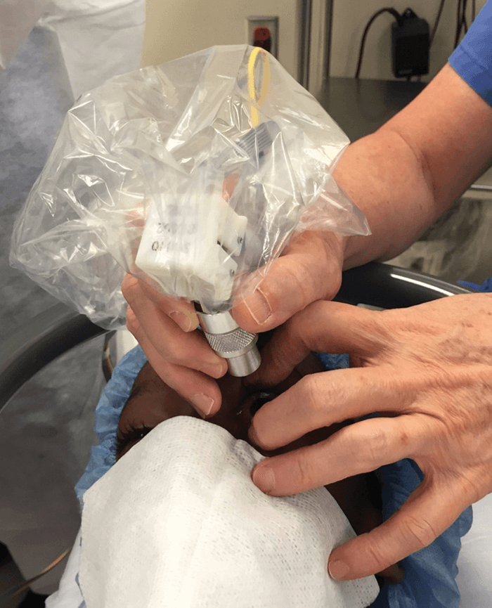
In a bid to overcome the challenges associated with acquiring retinal images from young children, and to increase image resolution, a team at Duke University have been developing an ultracompact handheld SLO/OCT probe (Figure 1). Having demonstrated  their device obtains high-resolution retinal images in children as young as 14 months of age (1), Cynthia Toth, Joseph Izatt, and Francesco LaRocca tell us more…

Why did you decide to develop a handheld SLO/OCT probe?

Measuring the impact of injury or diseases (genetic or otherwise) on the photoreceptors of infants has not been possible, because diagnostic tools that examine and image the retina – although well-designed for adults – are exceedingly difficult to use in infants and young children.

Some weigh several pounds, making holding them still over a child’s eye tiresome and difficult, and none provide a high enough resolution to see individual photoreceptors. Without the ability to image children’s eyes at high resolution, studying how our retinas grow and change during the crucial early stages of development is difficult. This limits our knowledge of how diseases affect a child’s vision early in life and makes diagnosis of blinding diseases that affect children more difficult.

What are the key findings so far?

The probe’s novel optical design and ability to image via both SLO (for high lateral resolution) and OCT (for high axial resolution) makes it uniquely optimized for imaging critical cell layers of the developing retina – like photoreceptors. For the first time, we have been able to evaluate the density of cone photoreceptors in infants during an eye examination under anesthesia (1).

We found that photoreceptor densities far away from the fovea for very young children were greater than those at the corresponding location for adults, supporting the hypothesis that there is a central ward migration of cones with age. Tests also showed different microscopic pathological structures in diseased children that are not normally visible with current lower-resolution clinical-grade handheld systems.

Figure 1. A clinician using the handheld probe to collect high-resolution SLO and OCT images of the retina from an anesthetized child. Credit: Joseph Izatt, Duke University.

Can you summarize how the probe is used?

Imaging sessions take approximately 10 minutes, including SLO imaging at two different fields of view (field of view [FOV]; 6.4° × 8.8° and 3° × 3°) and OCT volumetric imaging (6.4° × 6.4° FOV) for multiple regions of the retina near the fovea and optic disc. SLO and OCT can be switched by simply changing the fiber connections to the appropriate detection hardware.

Any challenges?

The traditional 4F correlator-based telescope design limited how small we could make a handheld probe, so we created a new design using converging rather than collimated light which reduced the telescoping length of the device by a third. However, this design inherently introduces significant optical aberrations (mainly field curvature) into the system, which we could not correct for with the few off-the-shelf lenses with high focusing power and small form factor that are available today. To enable high-resolution imaging at a minimal device size, we designed and specially fabricated custom lenses. Creating a custom mechanical design to hold these tiny optical components stably (whilst allowing room for focus correction adjustment) without adding much bulk or weight to the probe was another challenge.

What has been the main feedback so far?

Very positive – clinicians really appreciate imaging performance, and how light and compact the handheld probe is. Some enhancement requests include increasing FOV and incorporating motorized focus adjustment over a longer correction range to facilitate imaging of subjects with significant refractive error.

Next steps?

We will finish testing and developing the next generation probe, and prepare the process to make our device more widely available, which is anticipated within 3–5 years.

Achieving precise photoreceptor measurements in the retina opens doors to new research and tools that will be key in the future diagnosis and care of hereditary diseases.

Receive content, products, events as well as relevant industry updates from The Ophthalmologist and its sponsors.

When you click “Subscribe” we will email you a link, which you must click to verify the email address above and activate your subscription. If you do not receive this email, please contact us at [email protected].
If you wish to unsubscribe, you can update your preferences at any point.

  1. F LaRocca et al., “In vivo cellular resolution retinal imaging in infants and children using an ultracompact handheld probe”, Nat Photonics (2016). Advance online publication (doi: 10.1038/NPHOTON.2016.141).
About the Author
Ruth Steer
Related Case Studies
Business & Profession Glaucoma
TrabEx Pro: The Next Level in MIGS

| Contributed by MST

The Missing Piece of the Dry Eye Puzzle

| Contributed by Quidel

Uncovering Ocular Comorbidity

| Contributed by Quidel

Related Product Profiles
Uncover the Unique DNA of SPECTRALIS®

| Contributed by Heidelberg Engineering

Subspecialties Cornea / Ocular Surface
Tear Osmolarity – Empowering. Established. Essential.

| Contributed by TearLab

Subspecialties Retina
ForeseeHome® – remote monitoring to help detect wet AMD earlier and improve outcomes

| Contributed by Notal Vision

Product Profiles

Access our product directory to see the latest products and services from our industry partners

Here
Most Popular
Register to The Ophthalmologist

Register to access our FREE online portfolio, request the magazine in print and manage your preferences.

You will benefit from:
  • Unlimited access to ALL articles
  • News, interviews & opinions from leading industry experts
  • Receive print (and PDF) copies of The Ophthalmologist magazine

Register

Disclaimer

The Ophthalmologist website is intended solely for the eyes of healthcare professionals. Please confirm below: