The SPECTRALIS® Glaucoma Module Premium Edition (GMPE) is known for providing clinicians with a comprehensive toolkit to assess the structural loss characteristic of glaucoma. The multimodal approach empowers physicians to evaluate both routine glaucoma patients and those who present with a diagnostic conundrum. Now with deviation maps and Hood glaucoma report, as standard, it has never been simpler to offer accurate, objective glaucoma care, personalized to the patient – and the physician.
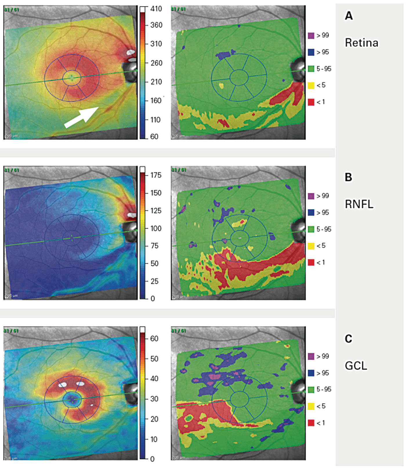
With the discrimination power of the SPECTRALIS GMPE deviation maps, clinicians can visualize structural loss at a glance. Comparison of thickness measurements with the reference database enhances their diagnostic value. The maps highlight the probability of measurements that are not within normal limits – revealing regions and associated patterns in specific retinal layers that have statistically significant thinner or thicker values. But that’s not all: the SPECTRALIS GMPE deviation maps also extend beyond the macula grid, delivering a wealth of additional diagnostic information.
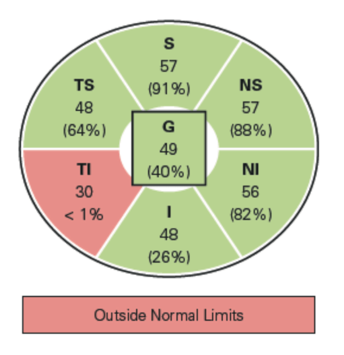
The thickness map color scales for the total retina and each retinal layer (retinal nerve fiber layer, ganglion cell layer and inner plexiform layer) have been optimized to offer a perceptually uniform representation of their anatomy, based on values taken from the reference database. The Anatomic Positioning System ensures accurate alignment of the six-sector grid with the reference database, according to the individual anatomy of each eye. The thickness maps also take into consideration the distance between the fovea and the center of Bruch’s membrane opening – adjustments that improve the accuracy and reliability of the deviations.
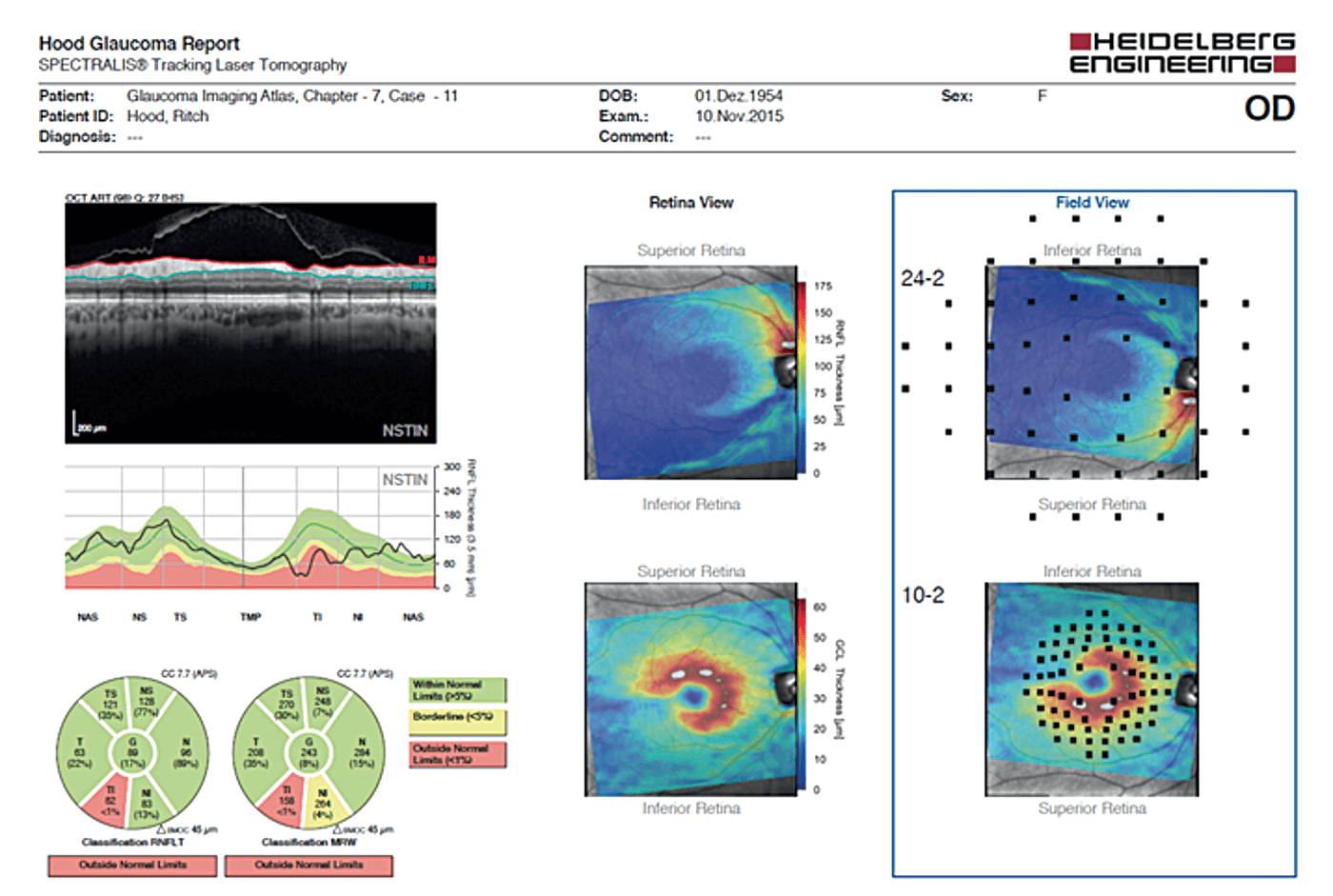
The Hood Glaucoma Report included in the SPECTRALIS GMPE combines and organizes the most pertinent OCT data from the ONH, RNFL and macula, empowering physicians to detect glaucomatous damage and relate information to visual field location in a clinically effective manner. More than that, the GMPE Hood Glaucoma Report presents RNFL data using an NSTIN RNFL profile, starting at the nasal-most point of the disc, before proceeding counterclockwise. This rearranges the RNFL profile so that thickness data associated with the temporal half of the disc is presented in the center. This NSTIN plot improves the utility of the RNFL information by optimizing its visual presentation in relation to visual field results. In addition, the large, high quality OCT image on the report confirms the details of the structural damage and the accuracy of the segmentation.
To intuitively compare structure and function, the RNFL and GCL thickness maps are rotated and overlaid with the visual field test points – 24-2 locations on the RNFL thickness map and 10-2 locations on the GCL thickness maps. The spatial agreement between the 10-2 and 24-2 visual field results and OCT thickness maps is impressive, especially when considering the various factors that can obscure this relationship – such as variability in anatomy and visual field testing among individuals.