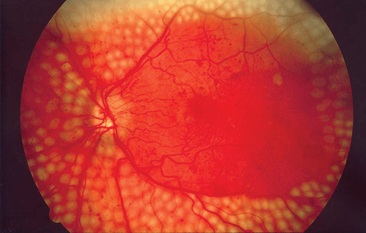
Retinopathy is a major concern among patients with diabetes – over a third of all such patients have a form of eye disease related to their condition (1), and the longer the time since diagnosis, the more likely a patient is to have diabetic retinopathy (2). The good news is that, with early diagnosis and treatment, the incidence and progression of retinopathy can be significantly reduced, and now, by using new ultrawide field (UWF) cameras, researchers at the Joslin Diabetes Center in Boston have uncovered lesions in the retinal periphery that may help predict patients’ risk of disease progression.
In the new study (3), Lloyd Paul Aiello’s group took a retrospective look at 68 eyes of 37 diabetic subjects, some of whom had a history of retinopathy and all of whom had undergone 200° UWF and UWF fluorescein angiography imaging. The researchers overlaid a template of the seven Early Treatment Diabetic Retinopathy Study (ETDRS) fields onto the images, then assessed them for the severity of the diabetic retinopathy, the presence of diabetic macular edema, and the presence of vascular abnormalities in the retina – if a lesion lay more than 50 percent outside the ETDRS fields, it was considered peripheral. The group found that such lesions were associated with higher nonperfusion of the retina, as well as with a substantially increased risk of disease progression. Eyes with peripheral lesions had over three times greater risk of progression, and almost five times the risk of progression to proliferative diabetic retinopathy. “The importance of the retinal periphery has been recognized for a long time, but we didn’t have the technology to image it until recently,” says lead author Paolo Silva. “With UWF, we’re able to see 82 percent of the retina in a single 200⁰ retinal image, with high resolution.” This presents a substantial advantage over the approximately 30 percent of the retina seen in standard ETDRS retinal imaging. The researchers also used stereographic projections to overcome the distortion normally seen in UWF images, allowing them to more accurately quantify the sizes of lesions and nonperfused areas. Of course, the Aiello group’s results still need further verification. In fact, the Diabetic Retinopathy Clinical Research Network is currently running a similar UWF imaging trial that involves more than 350 patients with diabetes. If the results of the larger trial confirm those of Aiello’s group, then it’s tempting to speculate that as UWF imaging technology decreases in price (and size) then it might supplant some of the current imaging methods as the first choice for diagnosing, staging and monitoring diabetic retinopathy.
References
- JW Yau, et al., “Global prevalence and major risk factors of diabetic retinopathy”, Diabetes Care, 35, 556–564 (2012). PMID: 22301125. RJ Tapp, et al., “The prevalence of and factors associated with diabetic retinopathy in the Australian population”, Diabetes Care, 26, 1731–1737 (2003). PMID: 12766102. PS Silva, et al., “Diabetic retinopathy severity and peripheral lesions are associated with nonperfusion on ultrawide field angiography”, Ophthalmology, [Epub ahead of print] (2015). PMID: 26350546.
