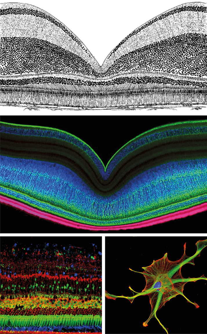
Imaging the cellular composition of retina back in the early 20th century required great patience and tenacity. The great advances in microscopy and photography were still to come, and it was the artistic ability of the scientists that determined how accurately anatomical structures were recorded (Figure 1, top).
Much like humans and some primates, certain fish and reptile species and birds possess an area of high visual acuity or even a fovea centralis. The upper picture shows a historical drawing from a histological cross-section of the central fovea of the European blackbird (1). The middle picture demonstrates multicolor immunohistochemistry staining, visualized by confocal laser scanning microscopy, of a cross-section at the edge of the fovea centralis of the European blackbird (2). The Müller glial cells of the retina are labelled in green (glutamine synthetase, a specific glial cell marker enzyme), the cone photoreceptor outer segments in pink, and the nuclei of the neurons in blue.
The bottom left picture shows immunofluorescent staining of calcium binding proteins in a cross-section from central retina of Macaca fascicularis, the crab-eating macaque (2). Antibodies against calretinin (blue) label the amacrine cells and rod outer segments. Antibodies against calbindin (green) label the cone photoreceptors and bipolar cells. Müller glial cells and retinal pigment epithelial cells are immunopositive for cellular retinaldehyde binding protein (CRALBP, red)
The bottom right picture shows a cultured Müller glial cell from an isolated human retina. The cell is stained with antibodies against interleukin 8 (red, an inflammatory protein produced by Müller glial cells during various retina pathologies) and against glial fibrillary acidic protein (GFAP, green) a glial cell marker (3). The cell nucleus is stained with Hoechst dye.
The last decades of the century saw the rise of immunohistochemistry and laser scanning microscopy, which enabled scientists to describe the molecular characteristics of cells at the protein level. Immunohistochemistry uses antibodies coupled with various fluorescent dyes to detect the distribution of marker molecules in cells and tissues. The distribution of the specific signaling or structural molecules can be visualized by laser scanning microscopy via wavelength-specific excitation of antibody-bound fluorescent dyes. Multiple labeling with different antibodies coupled with different fluorescent dyes in an individual tissue specimen, in combination with a laser scanning microscope, provide impressive colored images and allow conclusions about the particular combination of structural and signaling molecules in a given structure. Figure 1 (top and middle) provides an example where immunohistochemistry gives additional insights into the structural organization – and its consequences for visual sensory perception – of the fovea in the European blackbird, compared with a histological drawing. Furthermore, different neuronal cell types can be distinguished using the technique by their expression of specific marker molecules, and therefore, they can be characterized as a specific cell population with common molecular properties (Figure 1, bottom left). Even in cell cultures, where cells have changed their morphology and many biochemical properties, some marker molecules can be used to identify the origin of the cells (Figure 1, bottom right). Thus, advanced staining technologies and new microscopy techniques enable us to produce breathtaking cellular images and give us more fascinating insights in the beautiful organization of life.
Nicole Körber is a doctoral candidate at the Paul Flechsig Institute for Brain Research, Leipzig University, Germany. Peter Wiedemann is director of the Department of Ophthalmology at Leipzig University, secretary-general of Academia Ophthalmologica Internationalis and treasurer of the International Council of Ophthalmology. Andreas Reichenbach is acting director of the the Paul Flechsig Institute for Brain Research, Leipzig University, as well as head of the Department of Pathophysiology of Glia. Mike Francke is the leader of an independent research group at the Translational Center for Regenerative Medicine at Leipzig University.
References
- H Oehme, “Das Auge von Mauersegler, Star und Amsel”, J Ornithol, 103, 187–212 (1962). N Körber and M Francke, “The Winners of the Picture Competition 2012”, (2012). Available at: http://bit.ly/17P34Hp. Accessed January 16, 2015. M Francke, “Lange Nacht der Wissenschaften 2012”, (2012). Available at: http://bit.ly/1wfZbzU. Accessed January 16, 2015.
