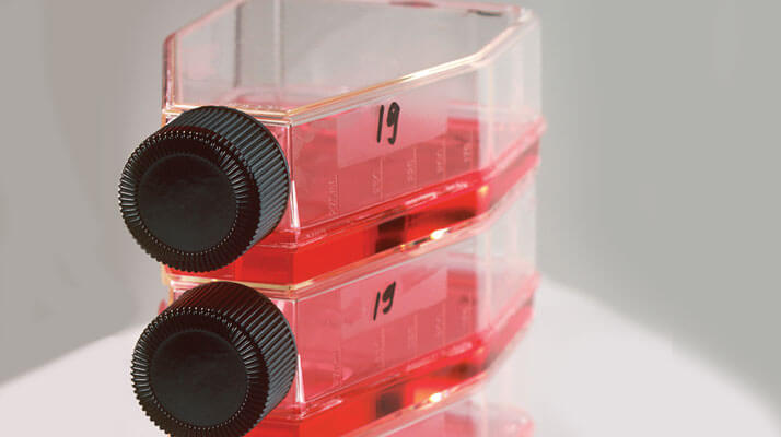
The retinal pigment epithelium (RPE) is one of the hardest-working tissues in the eye (if not the body), responsible for a multitude of functions, including nourishing the photoreceptors, elimination of metabolites, trophic factor production, the storage of retinal, and maintaining the blood-retinal barrier (1). It’s also a tissue that’s implicated in a multitude of disorders ranging from the well-known (like age-related macular degeneration) to the obscure (like monogenic retinal dystrophies and metabolic disorders). However, there’s one big issue that’s hindering the ability of vision scientists from understanding the RPE better: the lack of an appropriate tissue culture model. This has meant that research involving RPE cell culture has mostly been performed using primary human fetal RPE cultures – a valuable model, as primary culture allows the retinal and choroidal sides of the monolayer to be manipulated and measured independently, and fetal human cells are close in function to the retina in its native state. One of the major drawbacks of using a fetal culture model is that it may not accurately represent the physiology of the adult retina. Researchers could overcome that obstacle by using adult human RPE cultures – but these tend to lose their native physiology over time, resembling fibroblasts more closely than epithelial cells.
So how can scientists in the cell culture room avoid that problem?
It turns out that not all adult human RPE cells are created equal. A particular subset can be reverted in vitro to a self-renewing, multipotent cell type known as the retinal pigment epithelial stem cell (RPESCs) (2). That discovery opened the door to developing an adult human RPE culture without some of the problems that have plagued previous attempts. A group of vision researchers from the United States have now developed a new tissue culture protocol for RPESCs, and have determined that the cells produced by their method preserve many key features of the adult RPE, including their morphology, electrophysiological properties, and gene and protein expression profiles (3). The researchers were even able to culture RPESCs from patients with macular degeneration, diabetic retinopathy and glaucoma, a success that may ultimately lead to “disease in a dish” models of various RPE-related disorders. It’s too early to declare RPESCs the proverbial “better mousetrap” of RPE culture models. But so far, the authors feel that the new cells are promising as a useful model for diseases and disorders of the adult human RPE.References
- JR Sparrow, et al., “The retinal pigment epithelium in health and disease”, Curr Mol Med, 10, 802–823 (2010). PMID: 21091424. E Salero, et al., “Adult human RPE can be activated into a multipotent stem cell that produces mesenchymal derivatives”, Cell Stem Cell, 10, 88–95 (2012). PMID: 22226358. TA Blenkinsop, et al., “Human adult retinal pigment epithelial stem cell-derived RPE monolayers exhibit key physiological characteristics of native tissue,” Invest Ophthalmol Vis Sci, 56, 7085–7099 (2015). PMID: 26540654.
