Conexiant
Login
  • Corneal Physician
  • Glaucoma Physician
  • New Retinal Physician
  • Ophthalmology Management
  • Ophthalmic Professional
  • Presbyopia Physician
  • Retinal Physician
The Ophthalmologist
  • Explore

    Explore

    • Latest
    • Insights
    • Case Studies
    • Opinion & Personal Narratives
    • Research & Innovations
    • Product Profiles

    Featured Topics

    • Anterior Segment
    • Glaucoma
    • Retina

    Issues

    • Latest Issue
    • Archive
  • Subspecialties
    • Cataract
    • Cornea
    • Glaucoma
    • Neuro-ophthalmology
    • Oculoplastics
    • Pediatric
    • Retina
  • Business

    Business & Profession

    • Professional Development
    • Business and Entrepreneurship
    • Practice Management
    • Health Economics & Policy
  • Training & Education

    Career Development

    • Professional Development
    • Career Pathways

    Events

    • Webinars
    • Live Events
  • Events
    • Live Events
    • Webinars
  • Community

    People & Profiles

    • Power List
    • Voices in the Community
    • Authors & Contributors
  • Multimedia
    • Video
    • Podcasts
Subscribe
Subscribe

False

Advertisement
The Ophthalmologist / Issues / 2015 / Dec / Corneal Anatomy: Lightning Strikes Twice
Anterior Segment Cornea Research & Innovations

Corneal Anatomy: Lightning Strikes Twice

A specialized imaging technique has revealed two previously unknown structures of the cornea

12/4/2015 1 min read

Share

Figure 1. Two novel collagen structures were revealed by SHG imaging at the limbus: an anterior limbal cribriform layer and presumed anchoring fibers (1). Image credit: IOVS.

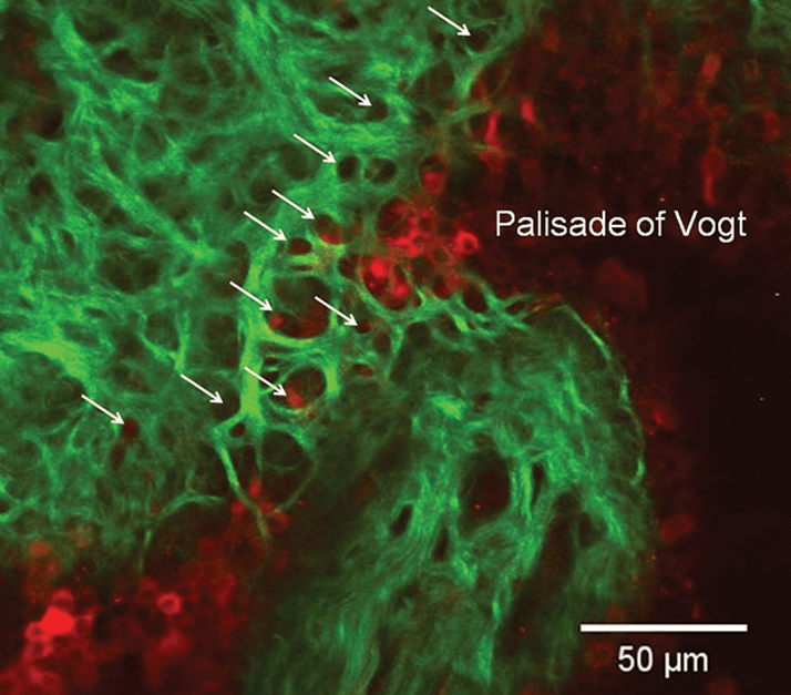
News that a new part of the body – a layer of the cornea – had been identified back in 2013 was such a big story that it transcended the world of biomedical news and hit the mainstream media. It’s happened again: another new part of the human anatomy has been discovered – and again, it’s in the cornea (1).Second harmonic generation imaging microscopy (SHIM) is an excellent method of producing extremely high-resolution images of certain (non-centrosymmetric) structures – one of which is collagen, the major structural component of the cornea. Researchers from the Albert Einstein College of Medicine in New York used SHIM on the cornea and managed to identify two new collagenous structures: a cribriform layer beyond the terminating loop of the limbal vasculature, and fibers connecting the peripheral cornea to the limbus (Figure 1). “What makes this finding extra interesting is the proximity of these new structures to a stem cell region where we already perform limbal stem cell transplants to replenish the corneal epithelium when it is lost to disease or injury,” said study co-author Roy Chuck.

The researchers were led in the right direction by several previous studies that used X-ray diffraction (XRD) imaging to identify collagen fibrils anchored in the limbal region. But XRD can only detect the presence of a given material, and not its architecture, whereas SHIM was able to resolve the conformation of collagen fibers, thereby revealing these previously unknown structures at the submicron scale in the eye – the anterior limbal cribriform layer and its presumed anchoring fibers. Although the function of these new structures remains unknown, the authors suggest a potential role in maintaining the stem cell and vascular surrounding environment in healthy corneas. The cribriform layer, which is composed of structural proteins like collagen and elastin, may also provide support to the region of the eye it underlies. “Hopefully we will be able to better understand the function of these newly discovered structures by monitoring their appearances in various disease states of the ocular surface,” said Chuck.

References

  1. CY Park, et al., “New details of the human corneal limbus revealed with second harmonic generation imaging”, Invest Ophthalmol Vis Sci, 56, 6058–6066 (2015). PMID: 26393473.

Related Content

Newsletters

Receive the latest Ophthalmology news, personalities, education, and career development – weekly to your inbox.

Newsletter Signup Image

False

Advertisement

False

Advertisement

Explore More in Ophthalmology

Dive deeper into the world of Ophthalmology. Explore the latest articles, case studies, expert insights, and groundbreaking research.

False

Advertisement
The Ophthalmologist
Subscribe

About

  • About Us
  • Work at Conexiant Europe
  • Terms and Conditions
  • Privacy Policy
  • Advertise With Us
  • Contact Us

Copyright © 2025 Texere Publishing Limited (trading as Conexiant), with registered number 08113419 whose registered office is at Booths No. 1, Booths Park, Chelford Road, Knutsford, England, WA16 8GS.

Disclaimer

The Ophthalmologist website is intended solely for the eyes of healthcare professionals. Please confirm below: