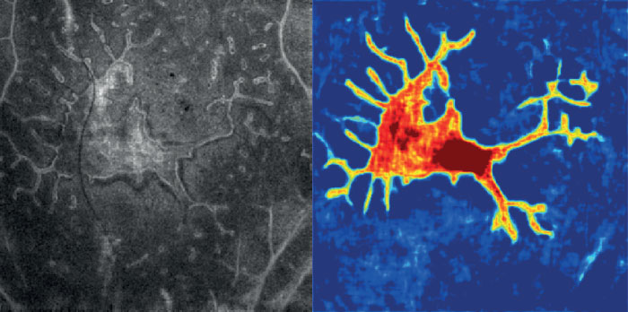
The image shows an en face OCT visualization of macular photoreceptor loss in choroideremia. Recently, en face OCT image processing methods together with machine and deep learning techniques have facilitated the segmentation of photoreceptor loss in diseases where low outer retinal layer contrast is used to yield to numerous segmentation errors and low-quality visualization of damaged areas (e.g. in inherited retinal diseases (1,2) and diabetic retinopathy (3)). Segmentation result by a fully convolutional network is also shown.
Credit: Acner Camino Benech, Oregon Health & Science University, Portland, USA
References
- A Camino et al., "Deep learning for the segmentation of preserved photoreceptors on en face optical coherence tomography in two inherited retinal diseases", Biomedical optics express, 9, 3092-3105 (2018). DOI:10.1364/BOE.9.003092. Z Wang et al., "Automated detection of preserved photoreceptor on optical coherence tomography in choroideremia based on machine learning", J. Biophotonics, 11 (2018). DOI: https://doi.org/10.1002/jbio.201700313. Z Wang et al., "Automated detection of photoreceptor disruption in mild diabetic retinopathy on volumetric optical coherence tomography", Biomedical optics express, 8, 5384-5398 (2017). DOI:10.1364/BOE.8.005384.
