Topcon Healthcare (Tokyo, Japan), a market-leader in the field of Optical Coherence Tomography (OCT) instrumentation, has consistently developed and innovated spectral domain (SD) and swept-source (SS) OCT technologies.
DRI OCT Triton represents the third-generation of OCT technology and combines deep scanning capabilities with the ability to penetrate media opacities with ultra-fast SS imaging (100,000 A-scans/s). Triton has the ability to capture comprehensive retinal and choroidal data in a single scan and produces more images to enhance the clinical decision-making process. In addition, Triton uses stereo photography to capture true-color fundus images in 3D, and thanks to its auto-mosaic panorama function, Triton provides wide coverage of the retina into the periphery (which includes most of the fundus).
In this article, world-leading retina and uveitis experts present cases from their practices. Utilizing the DRI Triton OCT and Triton OCTA was instrumental in efficiently diagnosing the ocular condition, assessing its severity, choosing the appropriate treatment, and monitoring progression.
Heloísa Nascimento, MD, PhD and João Rafael de Oliveira Dias, MD, PhD, Department of Ophthalmology, Federal University of São Paulo, Paulista Medical School – UNIFESP/EPM, Brazil
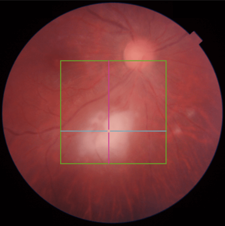
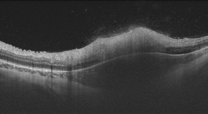
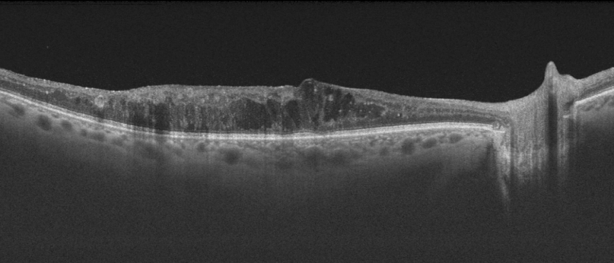
A 20-year-old female presented at our clinic complaining of floaters, photophobia, and ocular pain. Examination revealed acute toxoplasmosis in her right eye. Fundus images showed a white focal retinitis with overlying vitreous inflammation, vasculitis, and hemorrhages (Figure 1a). OCT examination, despite vitreous opacity, showed posterior vitreous inflammation with intraretinal alterations and important focal choroidal thickening (Figure 1b).
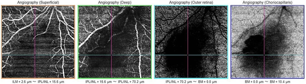
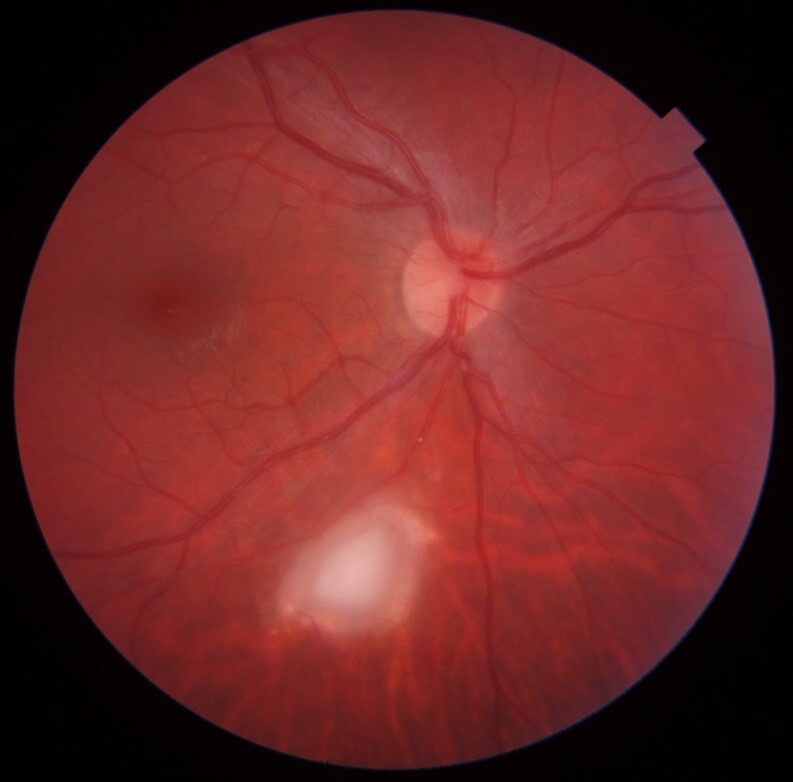
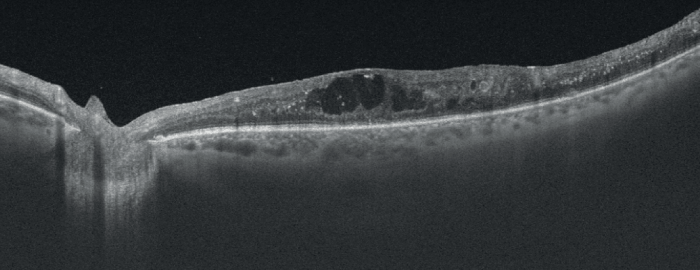
OCTA ruled out neovascularization and revealed focal areas of non-perfusion in the superior plexus (Figure 1c). These findings guided our treatment decisions for oral antibiotics and steroids. Five months later the inflammation had improved, the lesion had become well-delimited, and pigmentation had improved in the lesion borders with no signs of vasculitis (Figures 2a and b).
Follow-up OCTA indicated an improvement in retinal vascularization in the area adjacent to the lesion (Figure 2c).
Luis Arias Barquet, MD, PhD, Head of Retina Department, University Hospital of Bellvitge, Aggregate Professor of Ophthalmology, University of Barcelona, Spain
A 69-year-old Caucasian male presented with a 10-year history of diabetes, which was treated with oral medication and insulin. His diabetic retinopathy was previously treated with laser photocoagulation in both eyes (OU). He suffered progressive visual loss, OU for one month, with visual acuity of 20/50 (OD) and 20/80 (OS).
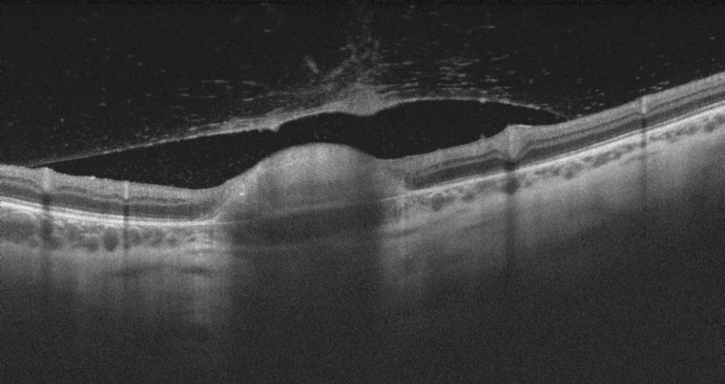
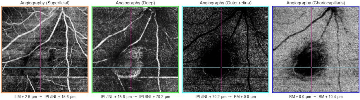
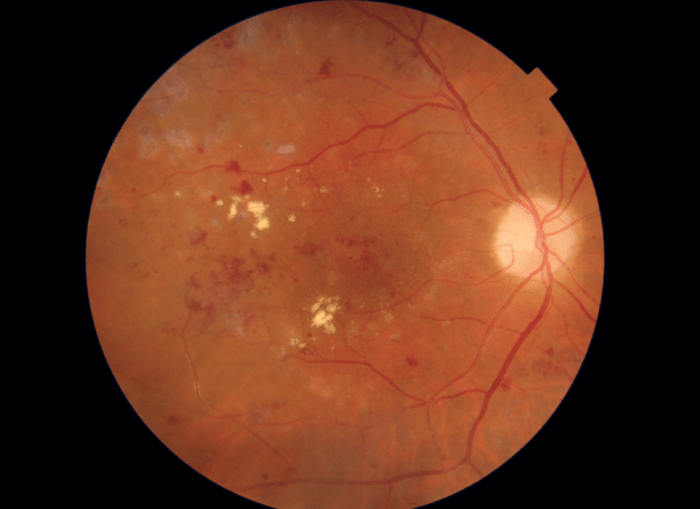
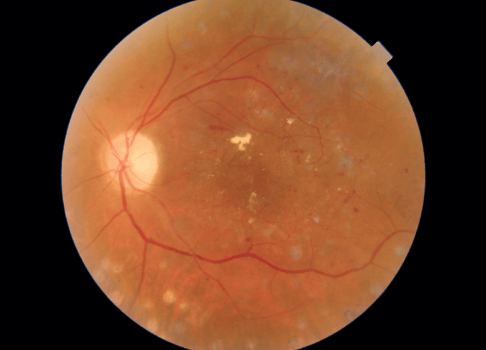
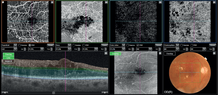
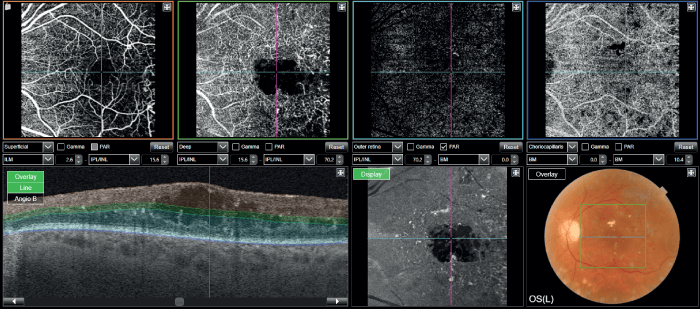
Fundus photography (Figure 3a: OD and 3b: OS) revealed pale optic discs, macular thickening with hard exudates, microaneurysms, and scars of the previous laser surgery, OU. OCT images revealed severe macular edema with large cysts, but relatively preserved outer retinal layer in OD (Figure 4a) and significant macular edema with large cysts, with compromised outer retina layers in OS (Figure 4b); discontinuity in the retinal layers may explain the degraded visual acuity, OS. OCTA superficial plexus evaluation, OD, (Figure 5a, orange frame) shows an enlargement of the foveal avascular zone, with capillary dilation, microaneurysms, and areas of peripheral ischemia. The dark areas in the deep plexus (Figure 5b, green frame) correspond to the inter-retinal cyst. The en face images (Figure 5a, middle lower image) help distinguish this from the presence of ischemia. OCTA evaluation, OS, (Figure 5b) provided similar information.
This case demonstrates the clinical advantages of utilizing multimodal imaging for patients suffering from diabetic retinopathy. Fundus photography is excellent for visualization of macular thickening, hard exudates, and blood. In contrast, OCT B-scans permit accurate assessment of the severity of the macular edema; they also support the evaluation of outer retinal layer integrity, which may inform visual prognosis. Finally, OCTA and en face imaging, enables the identification of ischemia, vascular alterations, and cysts. Such findings are essential for optimal management of patients with diabetic retinopathy.
It is now impossible to imagine a medical retina clinic without OCT capability. Among other OCT systems, the Triton is known to combine a user-friendly design with unmatched versatility. In addition, multimodal imaging of the ocular fundus is a great asset. The image acquisition panel allows the technician to focus on the patient and on acquiring good quality images, without distraction. Notably, the addition of OCTA to Triton has largely removed the need for fluorescein angiography in our unit. Finally, Triton’s anterior segment module has proven very useful in the assessment of a broader range of ocular conditions.
In my own experience, one of Triton’s most useful OCTA applications is for establishing the presence or absence of a choroidal neovascular membrane in patients with suspicious OCT images but no symptoms of visual loss or visual distortion. Furthermore, the uncertainty over whether to treat patients diagnosed with a newly identified entity of “non-exudative CNV,” when the finding is incidental and without symptoms or loss of vision, is ameliorated with Triton. In my practice, these patients are simply monitored with OCTA, which removes the need to expose the patient to repeated fluorescein angiography tests. This represents just one of several instances where Triton has significantly improved patient care.
Heloísa Nascimento, MD, PhD discusses real-life experiences with Topcon DRI OCT and OCTA Triton
I work at a private practice in Brazil, where I specialize in uveitis. I often have the opportunity to screen patients and, if there is an alteration, I perform more detailed examinations, such as OCT and OCTA. Those acquisitions can be challenging. For example, some patients have severe vitreous opacity, which complicates examination of the macula. I also have a high proportion of pediatric cases, and these patients do not always cooperate for fundus examinations.
In all these circumstances, Triton is very important as it allows me to see past most vitreous opacities to examine the retina, and it assists with pediatric fundus assessments because the acquisition is very fast. Having the right OCT system helps me perform all necessary examinations in a single appointment, which is always more convenient – and sometimes crucial – for the patient.
So, what is the right OCT system and how do we choose between the options? For me, quality is paramount. I don’t mind choosing a more expensive product if it is genuinely a better device which can enhance my clinical confidence. Practicality is another important factor. My husband and I, being retinal and uveitis specialists, respectively, must have a system that is reliable and simultaneously provides fundus photography, OCT, OCTA and angiography capabilities when needed. Familiarity is also desirable but not essential. One could say that I grew up with Topcon devices. My father, also an ophthalmologist, always used them in his practice. He appreciated their quality and simplicity and I expect the same, which is why most of the ophthalmology devices in my office are from Topcon!
For all these reasons, we chose the Triton OCT for our practice. Moreover, we believe that, if we are buying a device that we might use for many years, we should buy the best device possible – and for us, that is Triton. We also value Triton’s multimodality versatility and appreciate its simplicity (we find it easier to use than competing devices). The system is also fast and convenient, providing all the diagnostic tools I need in my own office, including true-color fundus photography, autofluorescence, angiography OCT, and OCTA. I have been using the Triton OCT for over five years and I can honestly say it helps my practice in a way others do not.
The Triton helps guide clinical decisions in my uveitis cases. Confident differentiation is very important for me because I see patients with a broad range of infectious and non-infectious diseases. If a patient has posterior uveitis, Triton OCT helps me to visualize whether it is in the retina or choroid. Similarly, in cases of acute retinal necrosis, the device helps me distinguish between causative agents such as: herpes, toxoplasmosis, or syphilis. In patients with cystoid macular edema, the Triton can support follow-up actions and can guide subsequent treatments, such as intraocular steroid injections. Therefore, it’s a great tool for differential diagnosis of various pathologies and uveitis entities, and it also assists with guiding my treatment decisions.
Interestingly, Triton is also appreciated by my patients as they like seeing the images it generates which I share with them digitally. In fact, I even share normal images with patients as it helps their confidence and makes them feel more secure.
I examine all my patients with the Triton mainly because I truly believe that OCT is the future, and it also helps me keep my patients happy and healthy.

Topcon Healthcare’s broad portfolio of SD and SS OCT applications, combined with continuous investment, allows the company to boast higher sales than any other manufacturer of OCT technology, accounting for almost 30 percent of total sales in this market segment (1). Topcon’s user-friendly offerings meet the constantly evolving needs of eye care professionals, ensuring a continued strong market lead for the years ahead.
These products are not available in all countries. Please check with your local distributor for availability in your country.
References
- Market Scope, “2020 Ophthalmic Diagnostic Equipment Market Report: A Global Analysis for 2019 to 2025” (2020).
