Legacy ultra-widefield retinal imaging systems require separate devices to capture images of the periphery on the one hand, and, on the other, high-definition color images of the optic nerve head. A far better option is to meld the two capabilities in a single instrument – as ZEISS has done with CLARUS 700 – so that clinicians can retrieve high-resolution pictures of the retina from macula to far periphery in one coordinated system.
True color
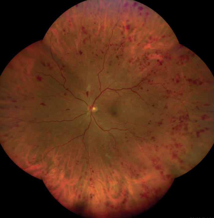
The CLARUS 700 is powered by Broad Line Technology: an innovative technology that illuminates only a small portion of the retina at any one time. This approach limits scatter and reflections, and thus enables the user to visualize high resolution details over a wide field. This is not possible with classic fundus cameras, which illuminate the whole field simultaneously. Furthermore, by sequential illumination with broad spectrum red, green and blue LEDs, Broad Line Technology captures images that closely resemble fundus colors as seen under direct clinical observation, thereby facilitating diagnostic analysis.
Comprehensive capabilities
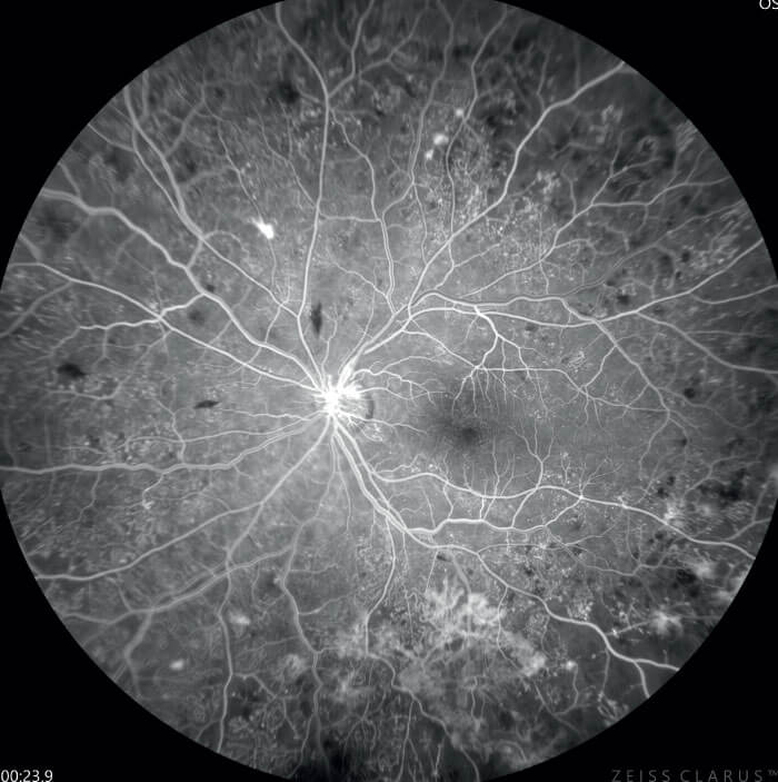
The recent addition of ultra-widefield high-resolution fluorescein angiography to the CLARUS capability set aids in visualization of vascular structures of the retina and the choroid. Fluorescein angiography may permit earlier identification of the more severe forms of diabetic retinopathy, and reveals non-proliferative diabetic retinopathy signals that may not be visible in color images. Beyond this, CLARUS 700 has a comprehensive suite of imaging modalities. These comprise of: infrared; red channel separation (reveals choroid in more detail); green channel separation (provides excellent contrast of retina, especially for vasculature and hemorrhages); blue channel separation (increases visibility of anterior retinal layer); FAF blue (helps reveal geographic atrophy); FAF green (can expose dry AMD); stereo imaging (for stereoscopic fundus evaluation); and views of the external eye. Together, these give the clinician unparalleled differential diagnosis capabilities, and may be employed together without compromising image quality.
Advanced features
CLARUS 700 utilizes algorithm-based technology to take the pain out of image processing:
• PrecisionFocus allows the clinician to rapidly focus in on areas of interest to reveal salient details without losing the macula focal point.
• AutoBright optimizes the brightness of angiogram images while preserving changes in signal intensity, relieving clinicians of the burden of image adjustment.
• GazePoint employs AI to determine the direction of the patient’s gaze without recourse to internal fixation
These features maximize workflow efficiency and decrease chair-time. In CLARUS 700, surgeons benefit from a single device that enables them to see more broadly, and make a faster, more accurate diagnosis.
Disclaimer: CLARUS 700 is 510(k) pending and not available in the US.