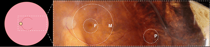
Part of the pathology of age-related macular degeneration (AMD) – and many other posterior segment diseases – is inflammation of the choroid and the accompanying retinal pigment epithelium (RPE). But it’s a patchy pathology; some regions are more susceptible to inflammation than others. North American researchers Jessica Skeie and Vinit Mahajan wanted to know why. Skeie and Mahajan are based in the Department of Ophthalmology and Visual Sciences’ “Omics” laboratory at the University of Iowa Carver College of Medicine. They had the means to identify the proteins across multiple regions of the choroid-RPE regions, the bioinformatical know-how to process the information, and three non-diseased eyes from the Iowa Lions Eye Bank. The plan was simple: take tissue samples from multiple regions of the choroid-RPE complex – the fovea, macula and the periphery Figure 1) – and create a map. So what map did they draw?
A molecular map that catalogued more than 4,000 unique proteins in each of the three areas examined, with differential regional expression patterns for almost 700 proteins that had previously been identified as risk factors for retinal diseases related to oxidative stress (1). Of note, the peripheral region contained unique antioxidant activity proteins, whereas many inflammation-related proteins and complement cascade activators were predominantly expressed in the fovea and macula regions sampled. One highlight was complement factor H (CFH). Certain CFH gene mutations can accelerate the development of AMD, and the study protein expression map revealed that CFH is most abundant in the fovea – the authors suggest that monitoring CFH expression in that region might act a marker of AMD disease status in certain experimental models. “This molecular map now gives us clues why certain areas of the choroid are more sensitive to certain diseases, as well as where to target therapies and why,” explained Mahajan. “Before this, we just didn’t know what was where. Now you can see all those differences that you couldn’t see before.” Previous studies have compared the abundance of single proteins in the fovea, macula, and periphery. The UI choroid-RPE map corroborates findings from these studies, but has also identified a treasure-trove of thousands more proteins that may be involved in vision loss. Mahajan likens it to a leap from the first topological drawings of a landscape to the detailed satellite images we have now.
References
- J.M. Skeie, V.B. Mahajan, “Proteomic Landscape of the Human Choroid-Retinal Pigment Epithelial Complex”, JAMA Ophthalmol. (2014) Epub ahead of print. doi: 10.1001/jamaophthalmol.2014.2065.
