It’s now more than half a century since David Hubel and Torsten Wiesel discovered that sewing a newborn kitten’s eye shut for the first three months of its life led to two things: full vision only developing in the open eye, and that this monocular deprivation leads to permanent electrophysiological and anatomical changes in the cat’s brain. Crucially, these changes were not seen if the eye was sewn shut closed after three months of age, so the immediate postnatal three months was named the “critical period” (or “sensitive period”) for vision development.
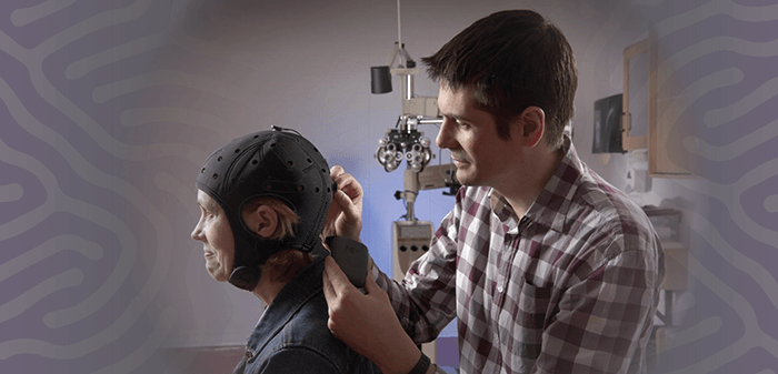
In humans too, the greatest effects of vision deprivation on the brain are immediately after birth – although at 24 months long, their critical period is considerably longer than in cats. The conventional wisdom was that it was essential that ocular abnormalities are identified and resolved during the first two years of life. Plasticity in the visual system progressively diminishes thereafter and appears to be almost gone by the age of eight – and therefore if diseases like amblyopia are not treated before eight years of age then the opportunity to save sight is completely lost. Today, we know that not to be the whole story – for example, children born blind with congenital cataract (and who were blind throughout the critical period, with cataract surgery occurring afterwards) can still experience significant improvements in vision (2). If you’re an adult with amblyopia, though, there’s new hope: transcranial direct current stimulation (tDCS). In the visual cortex, the structures that deal with inputs from both eyes are called ocular dominance columns (another Hubel and Wiesel discovery). The input from an amblyopic eye is subject to suppression from input from the fellow eye. This means that the amblyopic eye generates weaker visual evoked potentials (VEPs) than the fellow eye, and this is what leads to the characteristic visual deficits of the amblyopic eye, such as decreased visual acuity and impaired contrast sensitivity. It turns out that stronger suppression is associated with greater deficits in amblyopic eye contrast sensitivity and visual acuity. An international team of researchers based in Guangzhou in China, Waterloo and Montreal in Canada, Hong Kong, and Auckland, New Zealand decided to test whether noninvasive tDCS of the visual cortex would modulate VEP amplitude and therefore contrast sensitivity in adults with amblyopia (3). tDCS is an interesting approach – it can transiently alter cortical excitability and may even reduce suppressive neural interactions – such as from the dominant eye in people with amblyopia. Indeed, tDCS can be tuned – it appears to act in a polarity-specific manner; it has been shown previously that anodal (a-)tDCS of the occipital poles transiently decreases TMS phosphene thresholds, whereas the opposite effect is observed when cathodal (c)-tDCS is applied (4,5). The research team assembled 48 participants – 21 had amblyopia and 27 were present as controls. They received separate sessions of a-, c- and sham (s-) visual cortex tDCS. What they found was a-tDCS transiently and significantly increased VEP amplitudes in all eyes – amblyopic, fellow and control – and also contrast sensitivity for amblyopic and control eyes. c-tDCS decreased VEP amplitude and contrast sensitivity and s-tDCS had no effect. So what is behind these transient changes? Clearly, further work is needed to elucidate these mechanisms (and whether these changes can be made to stick), but the prime candidate for a-tDCS’ mechanism of action is reducing the amount of the inhibitory neurotransmitter, GABA, in the visual cortex, thereby reducing the chronic suppression of inputs from the amblyopic eye. a-tDCS also has excitatory effects, so the authors hypothesize that this “may lead to a transient enhancement of the cortical response to amblyopic eye inputs in the form of an increased VEP amplitude and improved contrast sensitivity.” Study co-author, Benjamin Thompson explained that these initial results demonstrate the proof-of-concept that will allow him and his research group to take the next step toward clinical trials, and that their “ultimate goal is to develop an evidence-based treatment that patients can receive right in their ophthalmologist’s office,” noting that, “We expect there are other primary visual cortex problems that we may be able to address with this method” – such as visual impairment secondary to stroke.
References
- T Wiesel, D Hubel, “Effects of visual deprivation on morphology and physiology of cells in the cats lateral geniculate body”, J Neurophysiol, 26, 978–993 (1963). A Kalia et al., “Development of pattern vision following early and extended blindness”, Proc Natl Acad Sci U S A, 111, 2035–2039 (201 PMID:24449865. Z Ding et al., “The effect of transcranial direct current stimulation on contrast sensitivity and visual evoked potential amplitude in adults with amblyopia”, Sci Rep, 14,6, 19280 (2016). PMID: 26763954. A Antal et al., “Manipulation of phosphene thresholds by transcranial direct current stimulation in man”, Exp Brain Res, 150, 375–378 (2003). PMID: 12698316. N Lang et al., “Bidirectional modulation of primary visual cortex excitability: a combined tDCS and rTMS study”, Invest Ophthalmol Vis Sci, 48, 5782–5787 (2007). PMID: 18055832.
