At this year’s ESCRS annual meeting, a group of high-profile refractive ophthalmologists were brought together in a special event organized by medical company CSO Italia. The aim was to connect researchers and clinicians with talks on the latest innovations in refractive techniques and technologies. The talks covered a range of topics from ground-breaking cross-linking strategies in keratoconus treatment to new imaging displays for the MS-39 OCT device, all delivered by world-leading experts.
Keratoconus concerns
Emilio Torres-Netto from the ELZA Institute and the University of Zurich, Switzerland, gave a talk on customizing cross-linking strategies in keratoconus – making the case that, just as techniques for cataract surgery depend on the specifics of individual cases, the same should be true of keratoconus treatment. Dr. Torres pointed out that the Dresden protocol is now around 20 years old and in some countries is still the only protocol used to crosslink keratoconic corneas. Given the condition’s complexities, cross- linking customization depends on available technologies, such as the EMAGine C-eye device.
The C-eye, with its integrated protocols, allows for treatment of very thin corneas with a stromal thickness lower than 400 microns. Torres explained that future developments with this device will allow for treating corneas with a new high- fluence protocol — just as effective as the Dresden protocol but much more rapid. Thanks to advanced penetration enhancers, it will also be possible to perform epi-on CXL treatments at the slit lamp. A combination of epi-on and epi-off treatment is involved in the new PACE (PTK-assisted customized epi-on CXL) technique that Torres-Netto and a group of researchers led by Farhad Hafezi are currently studying. In this new customized technique, utilizing the C-eye, the anterior segment OCT MS-39 and the Schwind Amaris 1050RS laser will allow not just for a stabilization of ectasia but also for an improvement of corneal topography.
This led naturally into Shady Awwad’s talk on wholistic keratoconus treatment. Awwad, who is Head of Cornea, Cataract and Refractive Surgery at the American University of Beirut Medical Center in Lebanon, pointed out that, when treated with penetrating keratoplasty, studies have shown that up to 27 percent of patients can end up with high irregular astigmatism, especially when the sutures are removed – so it’s essential to try alternative methods before turning to standard ablation or corneal transplant techniques. Furthermore, when ablation is used, it should be minimized to target higher-order aberrations only. After all, the more ablation is performed, the greater the stromal haze – impacting not only patients’ vision, but also their quality of life. Another important factor to consider is the extent to which patients might need repeat treatment. CSO Italia’s MS-39 AS-OCT’s ability to monitor corneal surfaces, along with the thickness of corneal layers and total corneal aberrations can be combined with the information of ocular aberrometry provided by the CSO Osiris-T aberrometer in order to offer a truly wholistic approach on the high order aberrations. This knowledge of total corneal wavefront along with the ocular wavefront, allows for surgeons to minimize corneal ablation up to the 44% in moderate keratoconus as detailed in research by Gore, Allan et al. (1)
In the future, the CSO MS-39 could be deployed alongside the Schwind Aramis 1050RS laser system allowing, for the first time, clinical treatment of the total corneal coma. Dr. Awwad also downplayed the advantages of mitomycin C, showing with patented OCT analysis software that its use can lead to much worse haze than treating without it. Awwad made the point that customized corneal ablation depends on detailed OCT imaging, mentioning the MS-39 specifically as a device that allows measurement of the total corneal wavefront.
Defocus data
Francesco Carones, Medical Director of the Carones Vision ADVALIA in Milan, Italy, offered a new approach to measure defocus curves. Defocus curves are an essential tool for a wide range of procedures, specially to test presbyopia correcting solutions, but they currently can be somewhat subjective - meaning they measure other issues than purely optics -and can be examinator dependent.
Dr. Carones presented a mathematical model to calculate an objective defocus curve from the wavefront derivatives measured by the Osiris aberrometer: he also demonstrated how this approach is more reliable in comparison to the solution based on the Wavefront map.
His approach also involved the use of the gPSF (Geometrical Point Spread Function) instead of the normal PSF and the use of new metrics to quantify the quality of vision. This opens the possibility of standardizing measurements according to different conditions, and gives the opportunity to describe patient visual performance after, for example, the implantation of a presbyopic IOL.
Carones then pointed out that this kind of objective measurement could develop to help clinicians choose the best IOL for implantation according to preoperative values such as corneal curvature, pupil size, and anterior chamber depth. Ultimately, surgeons could customize IOL selection based not on a theoretical or projected performance, but on an objectively measured and predictable performance tailored to the specific physiology of a given patient.
Essential equipment
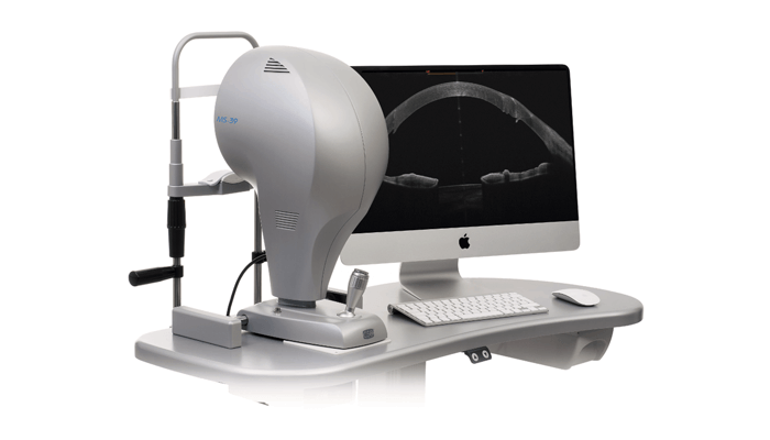
The first talk came from Dr. Dan Z. Reinstein of the London Vision Clinic, London, UK, and focused on displays for the MS-39. Reinstein demonstrated how summaries of four and six maps are set up in the London Vision Clinic in order to enhance and highlight topographic details. Given the ways in which treatment for keratoconus depends upon extremely detailed ocular mapping, the MS-39 that boasts dedicated displays for keratoconus was described by Reinstein as one of the most powerful tools for this disease management. Not only is this clinically useful, but it also makes good economic sense for doctors. The cost of the equipment is offset by the fact that greater accuracy and detail in diagnosis could make previously untreatable cases operable. As Reinstein put it, whatever outlay there is in terms of equipment, it is quickly paid off.
The final point described by Reinstein was how MS-39 can support the centration of SMILE refractive surgery thanks a new bilateral display that shows pupil offset simulating the position of the head of the patient in laying position; this unique feature is another way the MS-39 is an invaluable tool for all refractive surgeons. Reinstein’s final thoughts on the MS-39 make its advantages over other devices clear. CSO Italia, the company behind the device, has been in topography for 30 years, so its knowledge base and the software’s analytical capabilities are far ahead of its competitors. The MS-39 is “the slit lamp of the 21st century,” offering immediate cross-sectional imaging in the clinic. Its ability to offer high-resolution anterior segment imaging with Placido topography makes it indispensable for refractive surgeons and the detail of its stroma and epithelial measurements has become integral in developing new approaches to the treatment and management of keratoconus.
References
- Gore DM, et al., “Combined wavefront-guided transepithelial photorefractive keratectomy and corneal crosslinking for visual rehabilitation in moderate keratoconus,” J Cataract Refract Surg. 2018 May; 44(5) :571. PMID: 29891154.
