Powered by proprietary technologies like SSADA (split spectrum amplitude-decorrelation angiography), DualTrac™ Motion Correction and Projection Artifact Removal (PAR), Optovue’s newly developed high-density (HD) scan patterns provide best-in-class OCTA image quality for 6×6 mm macular and 4.5×4.5 mm optic disc scans. Known as AngioVueHD, sampling density has been increased from 304×304 A-scans to 400×400 A-scans. Overall sampling points have been increased by 73 percent, resulting in a 33 percent improvement in detail resolution compared to older OCTA scans. Now capillaries can be resolved to 15 µm allowing for visualization of very fine vasculature, which should provide greater confidence to physicians when imaging pathologies extending beyond the central 3×3 mm macular region. Importantly, the enhanced resolution is preserved over a wider field-of-view (FOV), so a 6×6 mm scan now provides the same detailed visualization of microvasculature and pathology as the previous 3×3 mm scan. This enables physicians to eliminate capturing two scans – one 3×3 mm scan for the best image quality, and one 6×6 mm scan for wide FOV. Now both high-resolution imaging and a wide FOV exist in one scan. Optovue currently offers the widest FOV and the highest density scanning of any commercial OCTA solution. AngioVueHD extends high-density imaging to montage mode by combining two high-density AngioVue scans to provide greater than 10×6 mm of coverage, while still visualizing fine retinal microvasculature details. AngioMontageHD automatically creates the image from scans of the macula and the optic disc, and is essential for imaging pathologies that extend into the periphery, such as diabetic retinopathy (DR) and retinal vein and artery occlusions. Beyond better image quality, high-density imaging is of paramount importance to the accuracy of OCTA quantification. Optovue’s AngioAnalytics, the world’s first OCTA quantification, leverages the improved resolution of AngioVueHD. Quantifying retinal vasculature provides an objective assessment of vascular traits more closely associated with visual functional outcomes than morphological traits alone. Current analysis tools include Flow Area in the outer retina and choroid (for assessment of choroidal neovascularization), Non-FlowArea of the superficial plexus (for assessment of ischemic regions in diabetic retinopathy) and Vessel Density analysis of the superficial plexus.
Maddalena Quaranta-El Mafoutouhi, MD, and Adil El Maftouhi, OD, Centre Rabelais, Lyon, France.
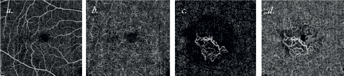
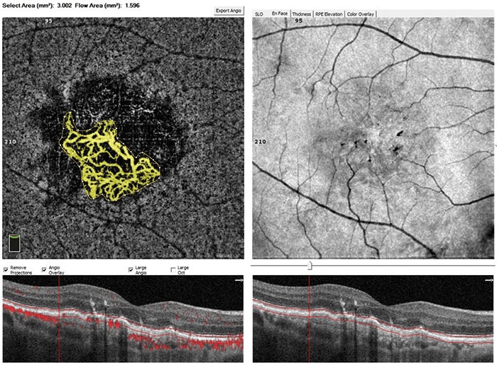
Here we present 6×6 mm AngioVue HD imaging of the retina of a wet AMD patient with type 1 (occult) CNV (Image 1), where the flow area was quantified using Optovue’s AngioAnalytics software (Image 2). The patient was followed-up for three months and underwent an induction phase of three intravitreal anti-VEGF injections. Their vision recovered from 6/10 to 10/10 on the day of the third injection – which is when these AngioVueHD 6×6 mm scans were performed. Furthermore, the new AngioVueHD software from Optovue now allows you to monitor CNV with a larger scan size of 6×6 mm, with similar details and vessel area quantification as 3x3 mm scans, but with the added benefit of a wider field of view.