Confocal imaging is well known in ophthalmology as the gold standard when it comes to high image quality. One key characteristic of confocality is rejecting or suppressing any
scattered/reflected light outside the focal plane – a feature that guarantees sharper images, better optical resolution, and greater contrast when compared with traditional fundus camera imaging.
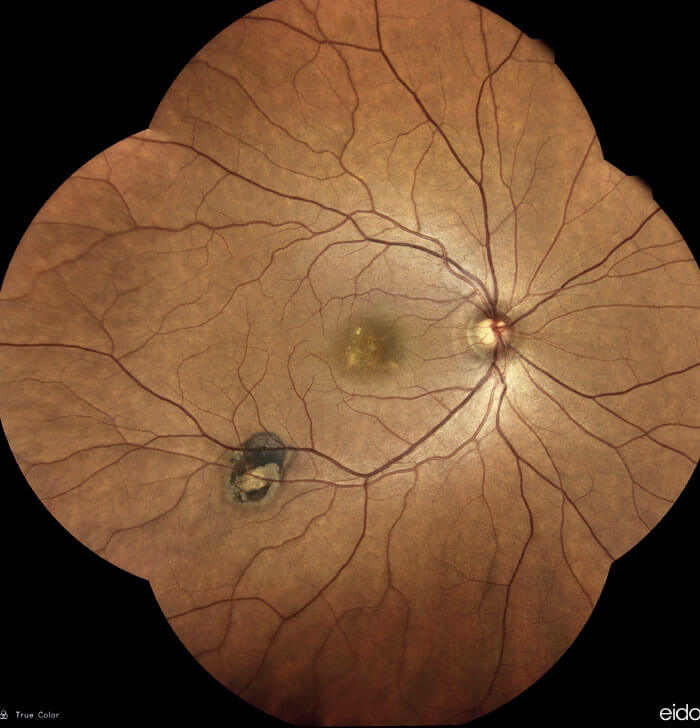
However, scanning laser ophthalmoscopy systems generally do not produce confocal images in true color; instead, they employ one or more monochromatic laser sources to image the retina, which results in grayscale or pseudo-color images. What makes CENTERVUE’s EIDON system unique is the white light LED source that uses the entire visible spectrum to illuminate the retina and capture fundus images. By creating retinal images in colors that are close to reality, ophthalmologists benefit from the most accurate perception of fundus anatomy. Moreover, the system allows specialists to work with very small pupils (down to 2.5 mm, without the need for dilation).
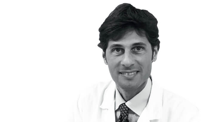
Giuseppe Querques, Associate Professor at University Vita-Salute, IRCCS Ospedale San Raffaele in Milan, Italy, is a retinal specialist, who treats patients with AMD, diabetic retinopathy, and other retinal vascular and genetic diseases. Querques has been using the EIDON system for the past few years – even from the prototype stage – and he believes that the EIDON system is a revolution in image quality. In particular, Querques is impressed with the clinical benefits that EIDON’s detail-rich images provide. “I think it is important to get TrueColor pictures for all my patients with macular and retinal diseases, as they guide treatment decisions. These days, having a sharp fundus image is vital; as we don’t tend to rely on dye angiography anymore, instead combining fundus imaging with OCT, it is the only way to properly document changes in the macula and retina, which aids the follow-up process, where even the slightest changes matter,” says Querques. “Looking at the crisp TrueColor image captured by EIDON is like having the real fundus right in front of me.”
Early detection of macular and retinal diseases often depends on distinguishing between normal features and potential signs of abnormalities. To Querques, this is where the detail rich TrueColor images are invaluable: “The breakthrough fundus imaging EIDON provides is vital in treating patients with AMD or diabetic retinopathy – with pathologies such as hemorrhages, micro aneurysms or microvascular abnormalities. TrueColor is a tremendous help in distinguishing between pigment and a hemorrhage – a crucial difference, which informs the potential AMD diagnosis.”
An important feature of the EIDON system is the ability to scan through cataracts and other media opacities. Querques, whose diabetic patients often suffer from cataracts, explains: “We have experienced major issues with other devices used for capturing fundus images as they rely only on blue light that cannot pass through cataracts. EIDON is an excellent tool for obtaining high-quality images when scanning through cataracts ahead of cataract surgery.”
Mosaic mode – EIDON’s wide-field capability of up to 150° – allows clinicians to see small details and changes as clearly in the (mid) periphery as in the central region, and with the same resolution and quality. According to Querques, wide-field imaging has changed retinal specialists’ perception of the diseases they treat, by awakening them to the crucial importance of picking up even the smallest abnormalities in the periphery. “Having a wide-field confocal TrueColor image is extremely important to me. With Mosaic, I know that I’m getting superior clarity, both in the center, and in the periphery. As EIDON is a fully-automated device, it is very easy to get the Mosaic images: once we have chosen the area of interest, the machine acquires the images automatically, making it a simple and quick process for the technician, the physician, and the patient.”
Indeed, the EIDON device works in fully-automated mode (including auto-alignment, auto-focus, auto-exposure and auto-shoot), but at the same time it provides flexibility of manual operation for custom image capturing. The device also exists in two other versions that can perform FAF (Fundus Autofluorescence and FA (Fluorescein Angiography).
Querques appreciates the system’s intuitive operation: “Using the device doesn’t take much time at all, so it makes the process a lot less stressful for the patient, and easier for the technician. In the past, to get the information on a diabetic patient’s condition, we had to take at least seven images of the retina, and the quality of them was often disappointing. Now, with EIDON TrueColor confocal capabilities, we can get an ultra high-resolution, wide-fiel image quickly and efficiently, using the fully-automated mode.”

