Featuring
Dan Z. Reinstein, MD, MA(Cantab), FRCSC, DABO, FRCOphth, FEBO
- London Vision Clinic, London, UK
- Columbia University Medical Center, New York, NY, USA
- Sorbonne Université, Paris, France
- Biomedical Science Research Institute, Ulster University, Coleraine, UK
Karel Van Keer, MD PhD FEBO
- University Hospitals Leuven
Juan G. Arbelaez, MD
- Muscat Eye Laser Center
Jorge L. Alió del Barrio MD, PhD, FEBOS-CR
- Cornea, Cataract and Refractive Surgery Unit, Vissum (Miranza Group), Alicante, Spain
- Associate Professor, Universidad Miguel Hernández, Alicante, Spain)
Farhad Hafezi, MD, PhD, FARVO
- University of Geneva, Switzerland: Professor, Medical Faculty
- University of Zurich, Switzerland: Research Group Leader
- University of Southern California (USC) Los Angeles, USA: Adjunct Clinical Professor of Ophthalmology
- Wenzhou Medical University, Wenzhou, China: Visiting Professor
- ELZA Institute AG, Dietikon, Switzerland: Medical Director
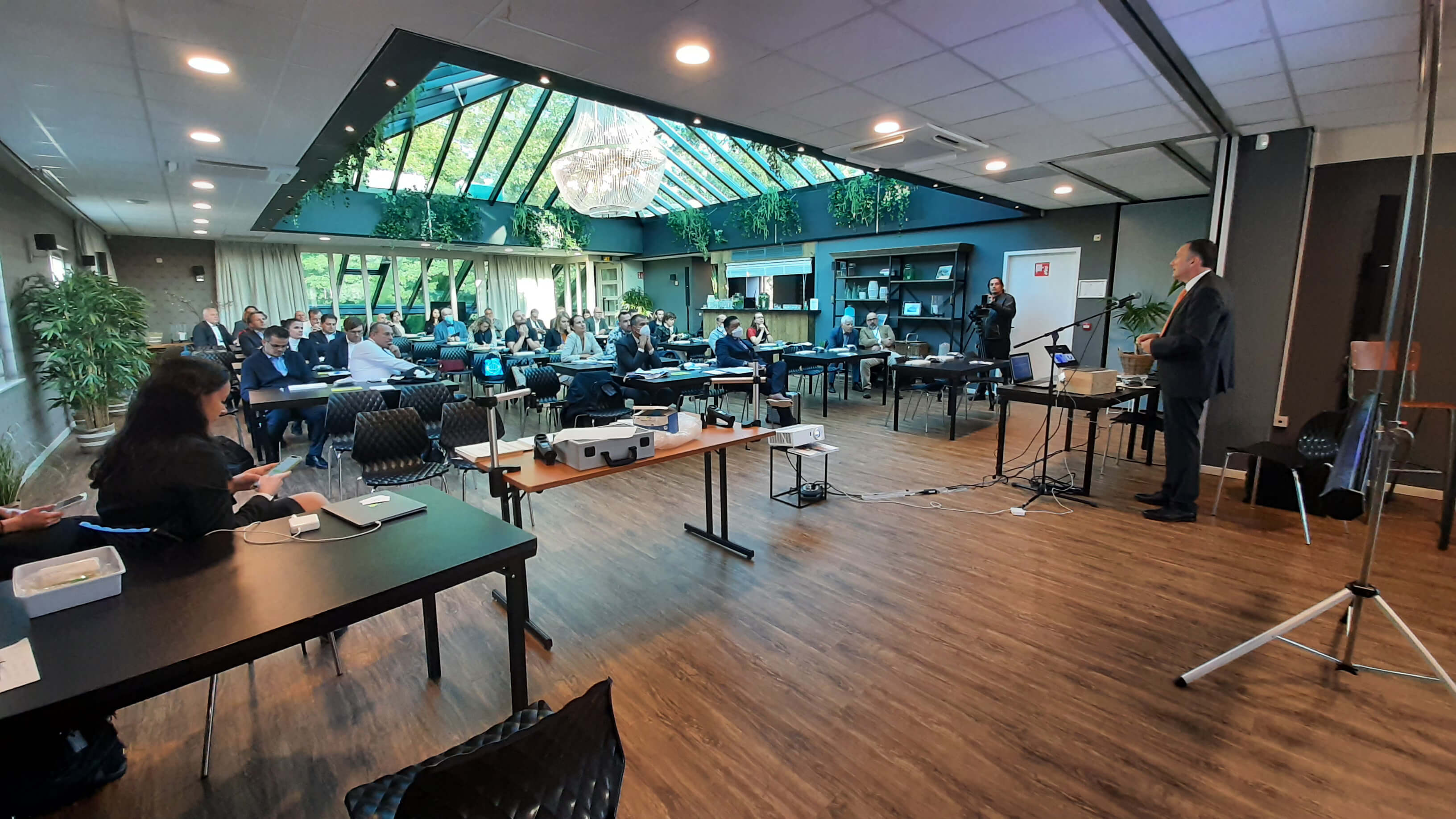
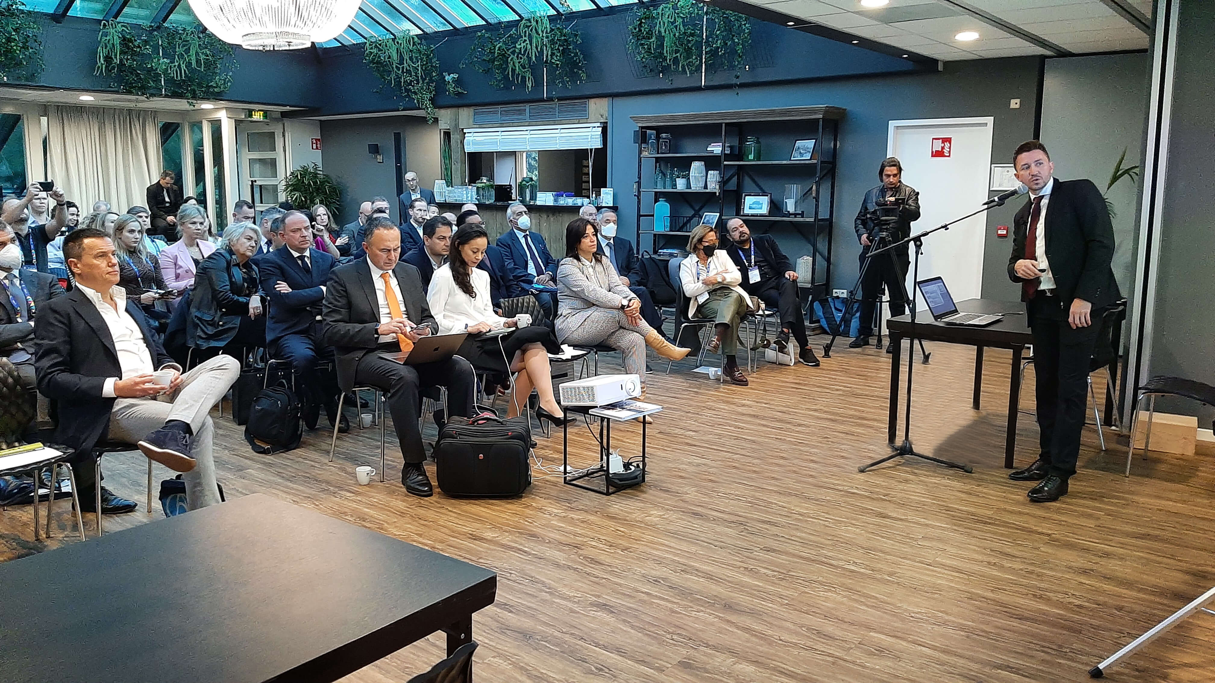
Combining Placido disk corneal topography and high-resolution OCT-based tomography of the anterior segment, MS-39 AS-OCT from CSO Italia boasts exceptional image clarity, advanced epithelial and stromal layer measurement capabilities, complete corneal aberrometry, and unparalleled keratoconus screening. MS-39 gives clinicians access to detailed data on pachymetry, elevation, curvature, and dioptric power of the corneal surface. Additionally, the device can offer IOL calculations based on Ray-Tracing techniques, pupil diameter measurements, and advanced tear film analysis. But how important are the above attributes to modern anterior segment specialists in their daily practice? In October 2021, five renowned experts explored this area in great detail.
Dan Reinstein’s presentation, “MS-39: The Slit Lamp of the 21st Century to Grow Your Refractive Practice,” highlighted the incredible progress made in the field of anterior segment diagnostics over the past two decades and the difference it has made to successful refractive surgery outcomes. He pointed out that this success depends on accurately selecting patients for refractive procedures based on the best available tools providing detailed anterior segment analysis. He recalled the moment a few years ago when he first saw MS-39 and realized that it marked the beginning of a new chapter in refractive surgery – the era of “superbly accurate” diagnostic system. For Reinstein, epithelial thickness profile is the crucial piece of information when assessing the cornea, and availability of epithelial mapping functionality of the MS-39 meant that once the device was commercialized, he immediately bought five units for all his testing rooms. Based on case studies from his clinical practice, Reinstein showed how measuring epithelial thickness and using the results to rule out keratoconus can help successfully qualify patients for LASIK – patients who would otherwise have been disqualified due to suspected keratoconus based on topography measurements alone (1).
Juan Guillermo Arbelaez presented findings in “Keratoconus Screening and Management and ICRS Treatment in the Era of AS-OCT.” Arbelaez compared sagittal and tangential curvature, defined on meridional basis and ignoring the orthogonal component of the curvature, with Gaussian curvature that doesn’t present that approximation and is independent of the axis, which makes it – in Arbelaez’s words – “extremely useful in defining ectasia.” Using various cases as illustrative examples, Arbelaez showed how MS-39 uses specific indices to identify issues with ectasia and other corneal weaknesses, utilizing anterior and posterior Gaussian maps, measurements of elevation, epithelial and stromal thickness to point to steeper, thinner or more elevated localized points on the cornea. He also mentioned a new symmetry index, which evaluates the central part of the cornea comparing it with the surrounding zone, and is therefore very useful in identifying cases of central keratoconus and helping with the condition’s correct classification.
In “ICL sizing made easy, repeatable and comfortable: the LASSO model,” Karel Van Keer talked about achieving best outcomes of Implantable Collamer Lens (ICL) surgery thanks to reliable data delivered by MS-39. As Van Keer explained, when it comes to safety in ICL surgery, one of the most important aspects is vaulting – finding the right space between the posterior surface of the ICL and the anterior lens capsule. If the vault is too low, it can lead to formation of cataracts, and if it is too high, it can reduce the aperture of the anterior chamber angle and lead to pressure-related issues. With ICLs available in four different sizes, it is crucial that the right size is chosen for the patient to achieve the perfect vault size of 500 µm – and this is where precise pre-operative data delivered by MS-39 come in. When the LASSO regression model to predict post-operative vaults was used, the data was extremely close to the desirable vault size (with maximum difference of just 2.61 µm) and the spread of the results was very narrow. Combining the precise measurements obtained with the MS-39 with the LASSO model has resulted in a formula that achieves better outcomes for ICL surgery patients.
Jorge L. Aliò del Barrio, in his presentation titled “Epithelium Mapping for Laser Refractive Surgery Evaluation,” explored the topic of modern corneal topography – in his words “an essential diagnostic tool for any ophthalmology clinic.” He highlighted the fact that modern devices combine various features – such as the latest Placido and AS-OCT technology, and many more – to deliver highest-quality data and measurements to aid anterior segment specialists’ diagnosis, pre-operative assessment and post-operative evaluation. According to Aliò, these features should include latest generation topography assessment, epithelial maps, ocular WF aberrometry, pupillography, HR AS-OCT (cornea, AC, angle, lens), lens biometry and IOL calculation, and dry eye assessment (like tear film analysis). Using various topography examples of refractive surgery candidates, such as corneal warpage, clinical and subclinical keratoconus, and pseudoectasia, Aliò asserted the value of accurate corneal epithelial and stromal thickness evaluation, supporting his statement with evidence (2, 3).
Leader in the keratoconus field, Farhad Hafezi, presented the C-eye: a portable, battery-powered UV irradiation device that allows clinicians to cross-link corneas both in the OR and at a wide variety of slit lamps, saving time and space and not requiring costly infrastructure. Hafezi covered a number of epi-off and epi-on protocols, showing the capabilities of the device with is capable of delivering both continuous and pulsed UV-A irradiation. As the cornea is thicker in the periphery, the irradiation profile of C-eye delivers more energy there than in the center of the cornea.
Whether it be keratoconus detection and diagnosis, selecting patients for refractive surgery or choosing the best ICL surgery parameters, MS-39’s multiple functions help leading anterior segment specialists better understand their patients’ visual issues and choose the most appropriate course of action to address them.
References
- DZ Reinstein et al., “Stability of LASIK in topographically suspect keratoconus confirmed non-keratoconic by Artemis VHF digital ultrasound epithelial thickness mapping: 1-year follow-up,” J Refract Surg, 25, 569 (2009). PMID: 19662913.
- A Vega-Estrada et al., “Corneal epithelial thickness intrasubject repeatability and its relation with visual limitation in keratoconus,” Am J Ophthalmol, 200, 255 (2019). PMID: 30689987.
- I Toprak et al., “Diagnostic value of corneal epithelial and stromal thickness distribution profiles in forme fruste keratoconus and subclinical keratoconus,” Cornea, 40, 61 (2021). PMID: 32769675.
