Subscribe to Newsletter
Subspecialties Basic & Translational Research, Neuro-ophthalmology, Retina

A Rare Discovery

The Campana cell (green) – spanning several layers of the retina from the photoreceptors to the retinal ganglion cells. John A. Moran Eye Center at the University of Utah.

The Campana cell (green) – spanning several layers of the retina from the photoreceptors to the retinal ganglion cells. John A. Moran Eye Center at the University of Utah.

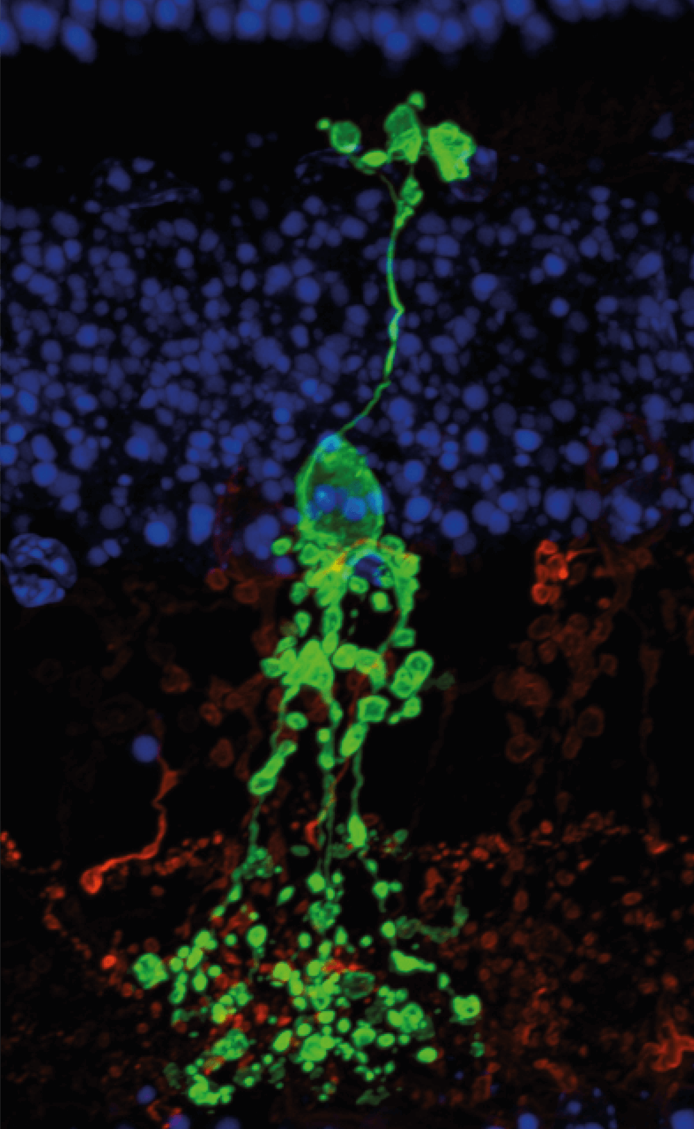
Over 100 years have passed since the identification of the five classes of retinal neurons. But in the adventurous world of science and ophthalmology, there’s always someone digging deeper. And now, researchers from the University of Utah’s John A. Moran Eye Center have revealed the existence of a retinal interneuron that doesn’t appear to belong to any of the existing retinal neuron classes. The team made the distinction based on the molecular properties, physiology, and morphology of the interneuron; indeed, its handbell shape gives the new Campana cell its name.

Though the Campana cell shares similarities with bipolar cells (relaying visual signals from photoreceptors to retinal ganglion cells) and shares features of amacrine cells (such as their short neurite morphology, some specific inhibitory signaling roles, and expression of certain biomarkers), they also differ significantly enough from these cells, according to the researchers, that they could be considered a new class of retinal neuron. And they may play an unconventional role in visual processing; for one thing, when stimulated with light, Campana cells react and activate for an abnormally long time – up to 30 seconds following a 10-ms stimulus. Lead researcher Ning Tian suggested the Campana cell may play a role in temporal memory, given that persistent firing of neurons in the brain is involved in memory and learning. Further research is needed to elucidate just what this uncanny cell class does – but we’ll be keeping our eyes peeled.

And then there were six

Here, for those of you with fuzzy physiology, we explore the other five retinal neuron classes – first described in detail by the great Santiago Ramón y Cajal in the late 19th century – and their specific roles in the amazing transformation of light into vision.

Photoreceptors

These light sensitive cells can be split into two sub-types – cones and rods. These enable us to see in color (cones) and in low levels of light (rods). These cells convert light into electrical signals that are transmitted through other retinal cells (one of these is the Campana cell) to get to the optic nerve.

Bipolar cells

The only cell type that connects to both the outer and inner retina – transmitting signals from photoreceptor to retinal ganglion cells. An additional layer of processing that is not seen in other sensory organs.

Retinal ganglion cells

Located near the inner surface of the retina, these cells relay the signals sent by both bipolar and amacrine cells to the brain. They have long axonal projections that form the optic nerve and allow them to form an information superhighway between the retina and the visual processing center.

Horizontal cells

These are regulators of the signals emitted from photoreceptors – providing important inhibitory feedback that enables increased contrast and adaptation to bright and dark light levels.

Amacrine cells

A cell type with over 30 different subtypes identified, so far… Named after their short processes (Greek for the etymologists amongst us), amacrine cells are another inhibitory and regulatory retinal cell class – interacting with retinal ganglion cells and bipolar cells.

Receive content, products, events as well as relevant industry updates from The Ophthalmologist and its sponsors.

When you click “Subscribe” we will email you a link, which you must click to verify the email address above and activate your subscription. If you do not receive this email, please contact us at [email protected].
If you wish to unsubscribe, you can update your preferences at any point.

  1. BK Young et al., Proc Natl Acad Sci USA, 118, e2104884118 (2021). PMID: 34702737.
About the Author
Geoffrey Potjewyd

Associate Editor, The Ophthalmologist

The lion’s share of my PhD was spent in the lab, and though I mostly enjoyed it (mostly), what I particularly liked was the opportunity to learn about the latest breakthroughs in research. Communicating science to a wider audience allows me to scratch that itch without working all week only to find my stem cell culture has given up the ghost on the Friday (I’m not bitter). Fortunately for me, it turns out writing is actually fun – so by working for Texere I get to do it every day, whilst still being an active member of the clinical and research community.

Product Profiles

Access our product directory to see the latest products and services from our industry partners

Here
Most Popular
Register to The Ophthalmologist

Register to access our FREE online portfolio, request the magazine in print and manage your preferences.

You will benefit from:
  • Unlimited access to ALL articles
  • News, interviews & opinions from leading industry experts
  • Receive print (and PDF) copies of The Ophthalmologist magazine

Register

Disclaimer

The Ophthalmologist website is intended solely for the eyes of healthcare professionals. Please confirm below: