- A decade ago, keratectasia treatment required heavy-duty surgery
- Today, most cases can be treated with corneal remodeling techniques in combination with refractive interventions
- The combination of intracorneal ring segments and phakic IOLs is a viable option for correcting corneal irregularities and large refractive errors in keratoconus patients
- The choice of technique(s) to use varies by a number of patient factors and the surgeon’s experience
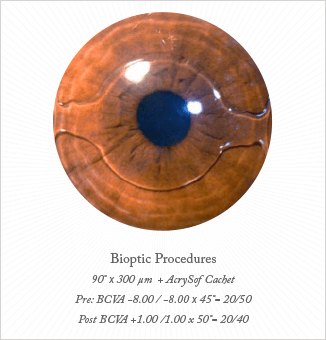
The efficacy of this sequential approach was illustrated by one of my patients who presented with pre-operative best corrected visual acuity (BCVA) of 20/50 and a refraction -8.00 sph -8.00 cyl × 45° in his left eye. I first implanted a 90° 300 µm ICRS and six months later implanted the pIOL. Six months after the second procedure, the patient’s refraction in that eye had improved to +1.00 sph -1.00 cyl × 50° and his BCVA was 20/40 (Figure 2).
The purpose of combining these procedures is twofold. One, the ICRS enables corneoplastic corneal remodeling: the surgeon remodels the cornea to improve the appearance of corneal topography. And two, to reduce astigmatism – without using invasive procedures such as penetrating keratoplasty or lamellar transplants. I tend to use ICLs or pIOLs to manage the high spherical error of refraction that is present in many patients with keratoconus.
Deciding Which Procedure to Use
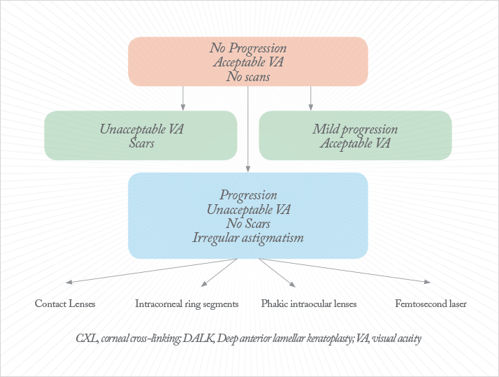
There’s a multitude of treatment options for keratoconus, and choosing the right one depends on any number of product and patient factors, and can be difficult. In my clinic, we follow a decision tree (Figure 1) to determine the best course of treatment. In patients with stable disease, no scars and an acceptable level of visual acuity (VA), we use contact lenses and spectacles. In patients with very low VA and scars, DALK is our preferred mode of treatment, and we can perform both traditional DALK and femtosecond laser-assisted DALK. We favor the use of corneal crosslinking in patients with mild disease progression and acceptable VA. For those with progressive disease with low VA and irregular astigmatism – but without scarring in the central cornea – we use contact lenses, ICRS, pIOLs, and laser treatment, either individually or in combination.
The last decade has seen a seismic shift in the treatment of keratectasia and keratoconus. Where spectacles, contact lenses, and penetrating keratoplasty for advanced keratectasia were the only treatment options available, today we have a variety of techniques at our disposal, including corneal crosslinking, intracorneal ring segments (ICRS), topography-guided photoablation, deep anterior lamellar keratoplasty (DALK), and phakic intraocular lenses/implantable collamer lens (pIOLs/ICL). Recently, I have started to combine two of these techniques, ICRS and ICL implantation. I have found that this creates a highly effective refractive intervention for the treatment of patients with keratoconus and high myopia.
I find that the combination of ICRS implantation with a pIOL provides a reliable keratoconus treatment option, while simultaneously correcting large refractive errors. To do this, I first implant the ICRS to control the keratectasia and reduce the cylindrical error; six months later, once the refractive results of the eye have stabilized, I implant a pIOL or ICL to correct the spherical error. One advantage of this approach is that it can predictably correct large spherical and cylindrical errors. Furthermore, patient recovery is a relatively speedy six months, compared to the 18-month recovery period that’s typically required after DALK surgery.
Although the same approaches used in eyes without ICRS can be used to calculate pIOL power after ICRS implantation, some special considerations are necessary in keratoconic eyes. We recommend that toric pIOLs are used in cases where the refractive and keratometric cylinders are in agreement and the axis of astigmatism from the refractive and keratoconus cylinders agree. In cases where the refractive and keratoconus cylinders diverge, we suggest treating only the high spherical myopic component of the refraction, hence we recommend that spherical IOLs are used. For any pIOL implantation, patients must have an anterior chamber depth of at least 3 mm and keratometry <55 D. If keratometry is >55 D, the IOL calculations become less predictable.

Clinical Experience
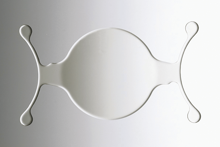
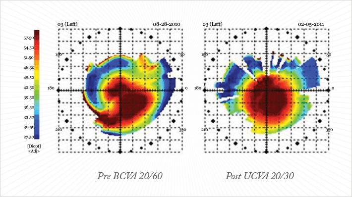
To determine the efficacy of this combination approach, I performed a study on the visual outcomes of keratoconus patients with large refractive errors who received the Keraring ICRS followed by implantation of the AcrySof CACHET anterior chamber pIOL. The patients included in the study had keratoconus, contact lens intolerance, clear corneas, maximum keratometry (K) reading <55.0 D, minimum K reading >40.0 D, and a minimum corneal thickness of 400 mm. All patients received the ICRS followed by an anterior chamber pIOL six months later. I implanted both the SI5 and SI6 Kerarings ICRSs in this study using the IntraLase femtosecond laser (Abbott LaboratoriesMedical Optics, USA) to create the tunnels for implantation. My rule of thumb with ICRS implantation is to have at least eighty percent corneal thickness at the site of implantation. In order to achieve better refractive results, I find it is necessary to make very tight tunnels. For the SI5, I use tunnels of inner diameter 4.8 mm and outer diameter 5.3 mm, whereas for the SI6, which is slightly larger, I use an inner diameter of 6.0 mm and an outer diameter of 6.8 mm and I make my incision on the steepest meridian (Figure 3). With the IntraLase, I manage to make precise tunnels within five to eight seconds.This study included 10 patients. The average pre-operative mean sphere in the group was -12.10 D ±4.48 D, (range -6.00 D to -21.50 D). The mean cylinder was -4.90 D ± 1.68 (range -2.50 D to -7.50 D); and spherical equivalent was -14.54 ± 4.25 (range -9.00 D to -24.00 D). Six-months after the pIOL was implanted, and 12 months post-operatively, mean sphere, cylinder, and spherical equivalent were 0.57 ± 0.91 D (range +2.00 D to -1.00 D), -1.23 ± 1.28 D (range -0.75 D to -2.50 D), and -0.15 ± 1.05 D (range +0.75 D to -2.25 D), respectively. The improvement in refractive results also led to an increase in visual acuity. Shown in Figure 4 is an example of a patient who had pre-operative refraction of -20.50 sph -4.50 cyl x 35° and pre-operative corrected distance visual acuity of 20/60. After the two procedures, his refractive results improved to -0.50 sph -0.75 cyl x 10° and uncorrected distance visual acuity improved to 20/30.

The last decade has seen a vast improvement in techniques for the treatment of keratectasia. Whereas penetrating keratoplasty and DALK used to be the most commonly used techniques for the treatment of keratectasia 10 years ago, today, these approaches are recommended only in advanced cases with central corneal scarring. The preference today is instead to remodel the diseased cornea using techniques such as corneal ICRS, crosslinking and thermokeratoplasty in combination with refractive procedures like pIOLs or customized photoablation. Indeed, the choice of modality used to treat any given patient will depend on a variety of factors, including patient parameters and surgeon’s experience. However, the combination of ICRS and pIOLs should be considered as a viable option for correcting corneal irregularity and large refractive errors in patients with keratoconus. Francisco Sánchez León is the Medical and Scientific Director of the Novavisión Laser Center in Naucalpan de Juárez, Mexico
