Ever since the first astigmatic keratotomy was performed over a century ago, ophthalmologists have known that changes in corneal biomechanics have refractive effects. Corneal collagen cross-linking (CXL) strengthens the cornea, and even it can be used for this purpose – for example, Avedro’s PiXL promises to correct refractive errors with selective cross-linking. But what’s next? Researchers from the Wellman Center for Photomedicine at Massachusetts General Hospital have revealed two-photon CXL (2P-CXL) (1). The paper’s corresponding author, Seok-Hyun (Andy) Yun explains, “In optical imaging, two-photon microscopy has become very popular, and we wanted to take advantage of these benefits and use them to improve conventional CXL, which is based on one-photon absorption of UV light.” In their study, the team selectively stiffened bovine corneal tissue in three dimensions using just riboflavin and 810 nm light pulses from femtosecond laser (1).
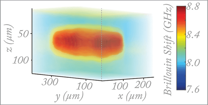
“Two-photon excitation confines tissue cross-linking to only where the laser is focused. This allows us to cross-link an arbitrary region deep inside tissue,” noted Yun, adding that “the degree of stiffening was similar to that with conventional CXL.” Central to the team’s research is in vivo Brillouin microscopy (2), which makes it possible to non-invasively visualize and characterize cross-linked regions. Yun explained, “Brillouin microscopy uses light to probe the mechanical properties of cells and tissues, and the principle relies on light scattering with spontaneous acoustic waves which are present in all materials.” Photons scattered by acoustic waves are sensitive to changes in compressibility and crosslink density, and local stiffening increases Brillouin frequency shifts, which are detected by high-resolution spectrometry (Figure 1). There is room for improvement: the procedure currently takes one hour to cross-link 1 mm2, but Yun believes that “this can be significantly improved with optimizations such as more efficient photosensitizers, laser pulses with higher peak powers, and optimized beam scanning patterns.” The team hopes to develop 2P-CXL for refractive error corrections in myopic and post-cataract patients, and Yun envisages an expedited regulatory approval: “One advantage is that many femtosecond lasers are already approved for use in the clinic, and the safety of riboflavin has been studied.”
References
- SJJ Kwok et al., “Selective two-photon collagen crosslinking in situ measured by Brillouin microscopy”, Optica, 3, 469–472 (2016). G Scarcelli, SH Yun, “Confocal Brillouin microscopy for three-dimensional mechanical imaging”, Nat Photonics, 9, 39–43 (2007). PMID: 19812712.
