Now in its 12th year, our Art of Ophthalmology feature continues to attract a wide range of submissions from across the art and medical worlds.
This year’s chosen submissions – sent in from medical professionals practicing in countries as diverse as Bangladesh, Poland, Germany, and Croatia – prove no exception, covering everything from the temporal impacts of glaucoma to the burning pain of inflammatory retinal diseases.
From more traditional analogue mediums of watercolor painting, photography, and mosaic, through to the more medically orientated slit lamp images and contemporary digitally created pieces, all of these submissions are united in their aim to present the viewer with insights into the ever-changing world of ophthalmology.
In these varying depictions of vision loss, ocular disease, and the inherent power of sight, we can gain insight into some of the powerful trends currently shaping the ophthalmic world – from the artistic perspectives of both long-term, experienced surgeons, as well as students and residents just starting out in their medical journeys.
Please find below the selection of our final chosen images. We hope these images inspire you as much as they did our editorial team.
AFTERIMAGE
Katherine McVeigh is a UK-trained ophthalmologist based in Berlin, with a subspecialty interest in oculoplastic surgery. Her creative work explores themes of perception and interpretation across textile and digital media.
“In ophthalmology, we are trained to identify structure within complexity, to recognize patterns, impose symmetry, and extract meaning from visual data. Diagnostic modalities such as OCT and fundus photography provide layered, often abstract representations that we interpret with clinical precision,” notes McVeigh. “This work, Afterimage, originates from photographs of clouds, forms that are inherently irregular, transient, and unstructured. Through layering and mirroring, these organic shapes are transformed into a symmetrical composition, reflecting how both the visual system and clinical practice impose order onto visual input. The image exists in the space between natural perception and constructed interpretation. It echoes the diagnostic process itself, finding coherence within ambiguity. While clinical imaging strives for objectivity, Afterimage reminds us that vision remains subjective, shaped as much by interpretation as by the image presented.”

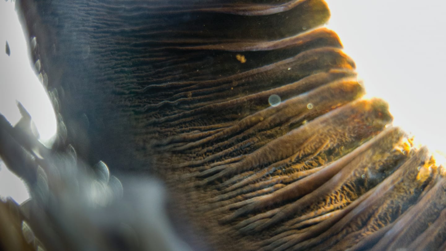
ARCTIC VOLCANO
Bartosz Ligęza earned his Doctor of Veterinary Medicine degree from Jagiellonian University, Poland, in 2018. From the beginning of his career, he has focused on veterinary ophthalmology, with a particular interest in ophthalmic photography and smartphone-based ophthalmic imaging. His work is based in Wrocław, Poland, at the Veterinary Ophthalmology Centre - EyeVet and Veterinary Clinic Novet. He manages an Instagram account, @vet_okulista, where he presents noteworthy cases.
Regarding the first of his two successful submissions, Ligęza explains how this image is “an ex-vivo picture taken with mirrorless camera (Sony a7r4a), macro lens with additional magnification backlit to highlight posterior uveal tissue.”

A DIVINE LOOK
Ligęza’s second submission, A Divine Look, is “an ex-vivo picture of a ciliary body and a lens taken from retina's perspective using a smartphone (Samsung Galaxy S24 ultra), an accessory macro lens and a flashlight.”
THE BURNING RETINA
Natasha Kesav is a physician specializing in medical retina and uveitis. Her clinical and academic interests focus on retinal imaging, inflammatory eye disease, and the intersection of systemic disease and ocular pathology. She is particularly drawn to the visual storytelling depicted in ophthalmic imaging. She hopes to highlight the aesthetic dimension of retinal disease while emphasizing the delicate complexity of the structures that make vision possible.
Of her submission, Kesav says: “The Burning Retina explores the visual and biological parallels between inflammation and fire. In inflammatory retinal diseases such as retinal vasculitis and uveitis, immune-mediated damage transforms delicate retinal vessels into sites of leakage, disruption, and destruction. In this piece, the retinal vasculature is reimagined as glowing rivers of lava radiating from the optic nerve head, evoking the intensity and unpredictability of an active inflammatory process.
“The fiery vascular network symbolizes both the destructive potential of uncontrolled immune activity and the striking beauty that can emerge from the microscopic landscapes of the eye. By blending clinical imagery with elements of natural phenomena, the work highlights how retinal imaging often resembles abstract art, revealing patterns that mirror those seen in nature.”

MARKET BLOOMS
Jaskarn Dhaliwal is a third year medical student at Queen's University, in Kingston, Ontario. She has a strong interest in a career in ophthalmology and equitable health care.
“This watercolor painting captures the quiet beauty of flowers that can brighten one’s day,” says Dhaliwal. “It shows a variety of blooms, highlighting how each flower, though different, has its own unique beauty. In both art and ophthalmology, paying attention to small details reveals complexity and beauty that might otherwise be missed. I hope to help patients continue to experience the beauty of the world by preserving their vision.”

TRICHROMATIC
Joanna McNulty is about to graduate from studying Medicine at Queen's University Belfast, Northern Ireland. She is also a practicing artist, having studied Art and Photography to Master’s level before embarking on her career in medicine.
“This photographic still life explores the principles of trichromatic vision through the interaction of red, green, and blue light,” explains McNulty. “Layers of colored acetate overlap to produce secondary hues, reflecting the process by which we construct color from the stimulation of three cone types. A cut image of an eye anchors the composition, introducing a human element within an otherwise abstract field. Suspended within shifting color, the eye becomes both subject and observer, suggesting the interplay between physiological function and lived perception. By reducing vision to its component channels and reconstructing it through layered light, the work reflects on the idea that sight is not a passive recording of the world, but an active, interpretive process.”

JOSHUA TREE
Michael P. Kelly has served as Director of Imaging at Duke Eye Center for 20 years as, and recently as Director of Remote Diagnostic Screening. He previously ran the imaging departments of West Coast Retina, California Pacific Medical Center, Cleveland Clinic’s Cole Eye Institute, and Cincinnati Eye Institute. He is a two-term past-President of the Ophthalmic Photographers’ Society, has 48 peer-reviewed publications, and has written seven chapters in four textbooks.
Of his submission, Kelly notes how "a career in ophthalmology has only deepened my fascination with the varied aspects of human perception – its reaches, its limitations, and its wonders."

EMBEDDED WITHIN
Faizan Naveed is a third-year medical student from Ontario, Canada, with an interest in both graphic design and ophthalmology.
“This piece,” Naveed says, “captures the eye as a reflection of both systemic disease and lived experience. The distorted inner world and reaching figure symbolize how ophthalmic pathology can become deeply embedded in one’s life, blurring the boundary between vision, identity, and lived experience.”

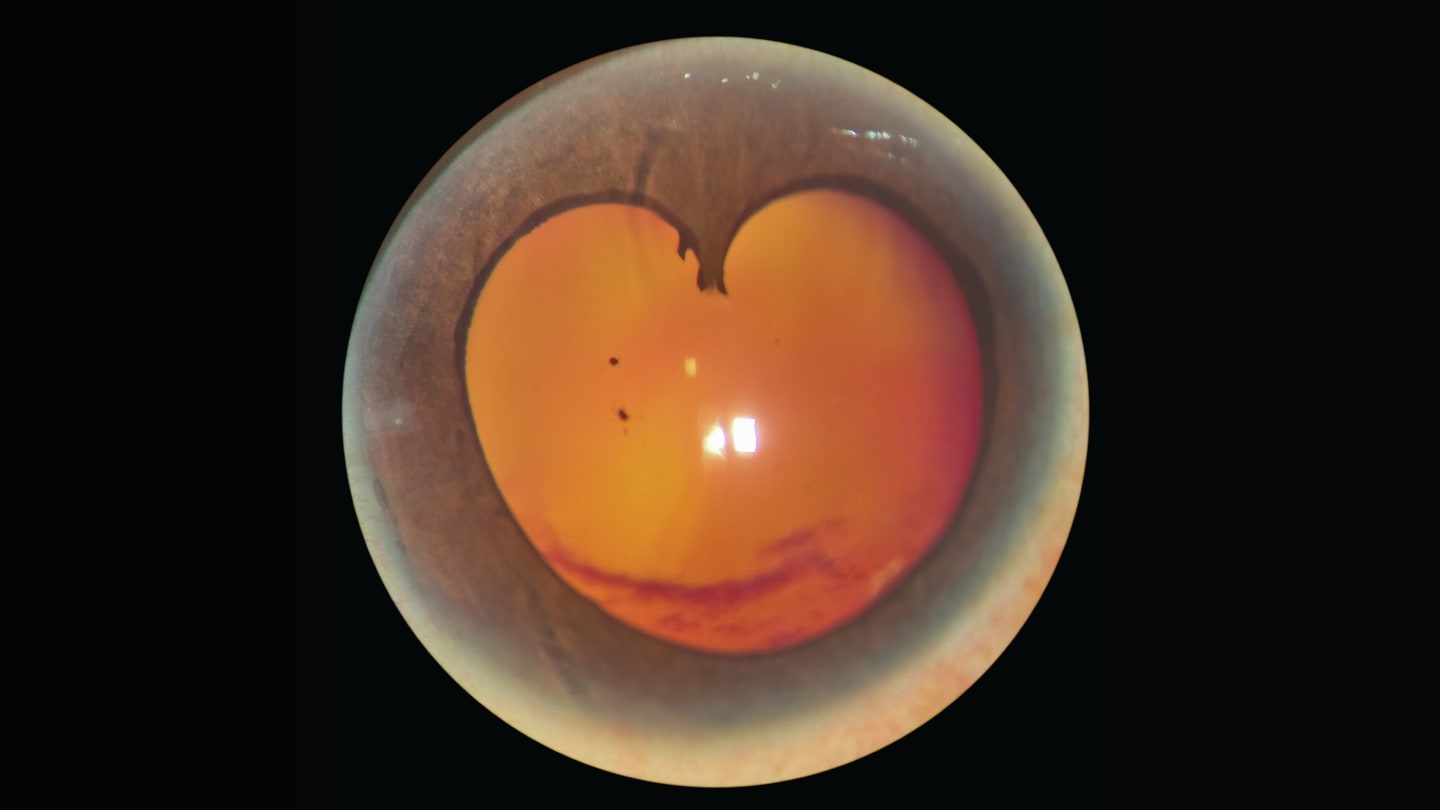
BLEEDING HEART, FADING VISION
Iftekher Iqbal is a glaucoma specialist from Bangladesh recognized for pioneering minimally invasive glaucoma surgery (MIGS) in the country and for managing complex glaucoma and cataract surgical cases in routine clinical practice. Working in a resource-limited setting, he focuses on practical surgical and clinical innovations that improve access to safe and effective glaucoma care. Beyond surgery, he has a keen interest in ophthalmic photography and digital imaging, capturing clinical moments that reflect both scientific precision and the quiet artistry of the human eye.
His first submission, Bleeding Heart, Fading Vision, is “a slit-lamp image capturing advanced diabetic eye disease with neovascular glaucoma. A heart-shaped posterior synechiae forms a striking central motif, with dense vitreous hemorrhage and a faint retinal detachment in the background. This single frame reflects the silent, progressive, and often devastating consequences of poorly controlled diabetes on the eye.”
SNAKEYE: THE LENS THAT SMILES BACK!
For his second image, Iqbal submitted “a slit-lamp image of a 12-year-old boy that revealed a developmental cataract forming a striking SnakEYE pattern – two symmetric lens opacities resembling serpent eyes with a subtle central smile-like configuration.”
Iqbal notes how “the image highlights how congenital lens changes can create vivid, almost expressive visual patterns while significantly affecting vision.”

TIME VERSUS GLAUCOMA
Tutut Nurjanah Sudiran is a medical doctor from Indonesia and a research associate at Bascom Palmer Eye Institute, focusing on glaucoma. Her work spans basic science and translational research. Alongside her passion for ophthalmology, Tutut creates visual art and immerses herself in music as a form of creative expression.
“The eye does not feel it happening,” Sudiran says of the glaucoma process she depicts in her Procreate image. “The clock does not stop; the loss of vision never comes back.”

FUNDAMENTALS OF OPHTHALMOLOGY
Borna Rupčić is training as an ophthalmology resident with hands-on experience in comprehensive eye care and patient-centered clinical practice at Županijska bolnica Čakovec in Croatia.
“As an ophthalmology resident who likes to express himself in various art forms,” he says, “I took inspiration from my day-to-day at the Eye Clinic and tried to put a fun artistic spin on fundus and refraction examinations. It was made on a 20 x 24 cm canvas using acrylic paint and highlighters.”

THE BEAUTY THROUGH THE LENS
Devina Ramesh is a third year medical student at Queen’s University, Canada, with an interest in ophthalmology. A self-taught artist working in a variety of mediums, mainly acrylic paints, she is drawn to the beauty of ocular anatomy and the personal stories that vision can hold.
“This piece is an abstract interpretation of the retina, created to capture the inherent beauty of ocular anatomy while preserving what makes it so visually striking,” Ramesh says of her submission. “ I wanted people to look at it and feel the beauty of the anatomy itself: the branching vessels, the glow of the fundus, and the vivid contrast of light against the dark background.
“What draws me to the retina is not only its function, but its visibility,” she adds. “Fundoscopic examination offers a rare privilege in medicine: the ability to non-invasively observe living anatomy in real time. It is a moment where structure, physiology, and art converge, where we are simply witnessing. With this painting, I wanted to turn that sense of awe into art and invite the viewer to see the retina not just as a site of diagnosis, but as something beautiful in its own right.”

RIYAZ’S VASES
“I have had the privilege to practice ophthalmology for over four decades and I feel proud to witness such huge advances in the field over this time,” says contributor Riyaz Ahmad. “As a young boy, long before picking up an ophthalmoscope, I picked up a paintbrush and palette. I have found painting to provide a sense of calm and focus, allowing me to think creatively – this has benefited my wellbeing and ultimately made me a better doctor.”
Regarding his submission, Ahmad says, “This watercolor painting is inspired by Rubin’s Vase, an optical illusion created in 1915 by Danish psychologist, Edgar Rubin. The viewer's brain can switch between seeing a central vase or two faces looking at each other. This version introduces the anatomical detail of the orbits and surrounding structures. The retinal vessels resemble branches of a tree so I have added skies and clouds, which are also visible above the frontal sinuses. In the central space, you can see a white vase and another vase bordered green. When viewed upside down, the painting reveals another bigger vase.”

FRAGMENTS OF SIGHT
Nesrine Rahmania is a Paris-based ophthalmologist affiliated with the Assistance Publique–Hôpitaux de Paris, specializing in cataract, corneal, and refractive surgery. She combines advanced surgical expertise with active involvement in research, and has published in peer-reviewed journals reflecting a strong commitment to advancing ophthalmic care.
Of her submitted image, Rahmania says, “This mosaic piece explores the human eye as both a biological structure and a poetic subject. Composed of meticulously assembled fragments of glass, the work mirrors the intricate construction of vision itself – where individual elements unite to form a coherent perception. The vibrant green iris, enriched with warm tonal variations, reflects the complexity of ocular anatomy and the subtle interplay of light and color. Through this process of fragmentation and recomposition, the piece evokes both the precision of ophthalmology and the emotional depth of seeing. This work bridges clinical insight and artistic expression, inviting the viewer to consider the eye not only as an organ, but as a gateway between the external world and inner experience.”

THE UNSEEN
Marie Jo Abdul-Hay is a medical student at the University of Ottawa in Ontario, Canada, with an interest in ophthalmology. She appreciates using art as a lens, to raise awareness on complex problems that require attention from the public and motivate collective solutions.
“Up to a quarter of people experiencing homelessness live with visual impairment, often from correctable causes like unaddressed refractive error. Mr Albert, a fictional character, is one of them. His foggy vision limits his independence and affects his ability to find a job,” Abdul-Hay says of her submission. “While homelessness is frequently framed through social and economic lenses, visual impairment remains a quieter obstacle. This comic strip aims to sensitize viewers to think of visual impairment as a functional necessity, not only a medical diagnosis. Despite the advancements in equity, and the fullness of life that people with blindness can have, many do not get the luxury of accessibility, whether that’s due to unawareness of resources, or inability to seek them. The medical field is growing towards clinics organized for marginalized populations, and Mr Albert was one of the lucky patients to get free glasses for his condition. Living on the streets, breaking possessions is sometimes inevitable, but as long as the glasses are functional, he is slightly better equipped to address some of the many problems that need to be solved to improve his quality of life.

