Neurotrophic keratitis (NK) has long been regarded as a disease of corneal denervation, with epithelial breakdown viewed largely as a downstream consequence of sensory loss. However, emerging evidence suggests this view may be incomplete. A new study published in the American Journal of Ophthalmology provides compelling in vivo evidence that limbal stem cell deficiency (LSCD) is not merely a late complication of NK, but a closely linked pathological process that evolves alongside nerve damage and immune dysregulation.
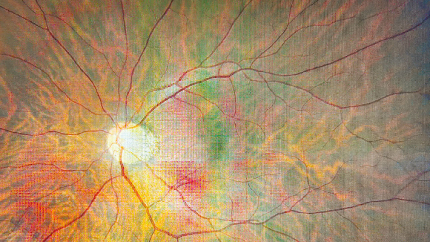
Using in vivo confocal microscopy (IVCM), researchers from the Department of Ophthalmology, Renmin Hospital of Wuhan University, Wuhan, China, examined corneal nerves, limbal epithelial stem cells (LESCs), and dendritic cells (DCs) in 54 patients with NK compared with 57 healthy controls. The findings reveal a striking triad of abnormalities: profound corneal nerve loss, marked depletion of LESCs across all limbal quadrants, and heightened immune activation.
Compared with controls, patients with NK showed dramatic reductions in corneal nerve fiber density, length, and branching, alongside a paradoxical increase in nerve fiber width – a pattern consistent with loss of fine branches and relative preservation of thicker nerve trunks. At the same time, LESC density was reduced by nearly 50% in all four limbal regions, indicating widespread stem cell compromise rather than focal damage. These structural changes correlated strongly with clinical severity, particularly fluorescein staining scores.
Immune cell activation emerged as a third key component. NK corneas exhibited significantly increased DC density, a higher proportion of activated DCs, and enlarged DC morphology – hallmarks of a chronically inflamed ocular surface. Importantly, DC density correlated negatively with both nerve parameters and LESC density, and positively with epithelial staining, suggesting that inflammation may actively contribute to both neural and epithelial failure rather than simply responding to tissue injury.
Correlation analyses reinforce the concept of a tightly coupled neuro-stem cell-immune axis. Corneal nerve fiber density was positively associated with the density of LESCs and negatively associated with fluorescein staining and DC density. In contrast, regional epithelial damage correlated more strongly with local LESC depletion than with nerve loss alone – a clinically relevant finding that underscores the importance of limbal health in persistent epithelial defects.
The study also provides valuable longitudinal insight into the effects of recombinant human nerve growth factor (rhNGF, cenegermin). Seven patients with stage 3-4 NK were followed after an eight-week course of topical rhNGF, with assessments extending to two years. Interestingly, early improvements were not neural but cellular and inflammatory: within four weeks, LESC density increased significantly and DC activation declined, accompanied by reduced corneal staining. Measurable nerve regeneration followed later, becoming apparent by eight weeks and more pronounced at two years.
This temporal sequence suggests that rhNGF may initiate corneal recovery by first stabilizing the limbal microenvironment and suppressing inflammation, thereby creating conditions that allow nerve regeneration and epithelial repair to occur.
While the improvements induced by rhNGF were sustained over two years, corneal nerve and LESC parameters did not fully normalize, highlighting that cenegermin promotes partial but meaningful restoration rather than cure. Nevertheless, the long-term persistence of benefit supports the idea that short-term intervention can trigger a regenerative feedback loop involving nerves, stem cells, and immune cells.
Taken together, this study reframes NK as a complex, multisystem ocular surface disease rather than a purely neurogenic disorder. It suggests that assessment and management of NK should extend beyond corneal sensation and epithelial integrity to include the limbal niche and inflammatory status. As therapeutic strategies evolve, preserving – or restoring – limbal stem cell health may prove just as critical as regenerating corneal nerves.
