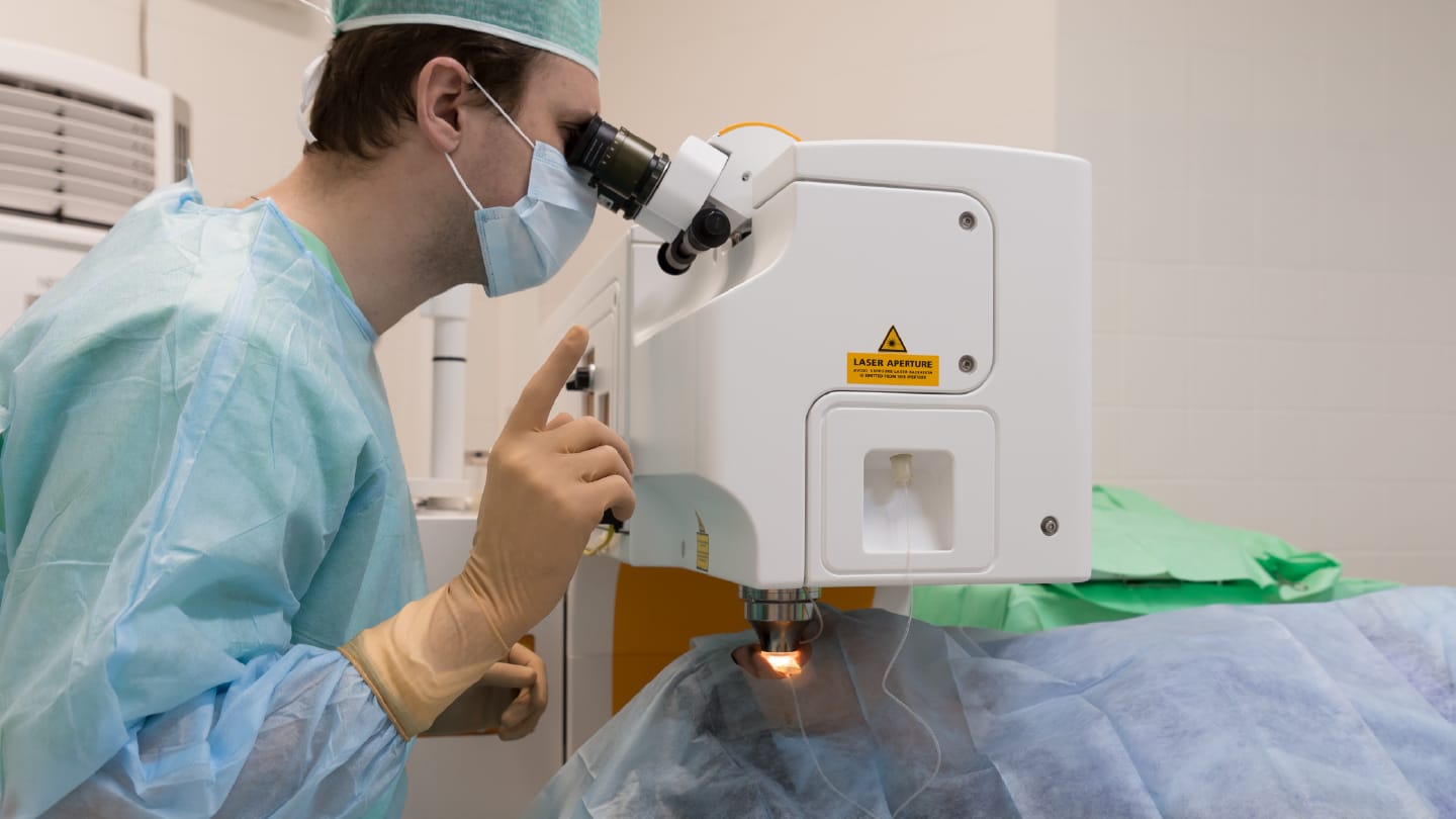
Achieving maximal tissue removal while sparing surrounding structures remains one of the central challenges of modern surgery. A new study published in Biomedical Optics Express has explored how deep ultraviolet (UV) ultrashort-pulsed lasers could offer an unprecedented level of precision for soft tissue ablation – potentially opening new surgical possibilities beyond their current use in ophthalmology.
While ultraviolet lasers are well established in corneal surgery, their use in other soft tissues has been limited. In this experimental study, researchers from the University of Edinburgh, UK, investigated how femtosecond laser pulses at a wavelength of 206 nm interact with ex vivo lamb liver, a soft, water-rich tissue chosen to model the challenges of delicate surgical environments (as well as mimic the less accessible brain tissue samples). By systematically varying laser pulse energy, spot size, and repetition rate, the team identified laser settings that enabled clean tissue removal with minimal collateral damage.
Using surface profilometry and histological analysis, the authors demonstrated axial ablation precision down to approximately 10 microns – significantly finer than that achievable with conventional surgical tools such as electrocautery, ultrasonic aspirators, or long-pulsed lasers. Importantly, they identified a clear “optimal ablation window” defined by laser fluence rather than energy alone. Within this window, tissue was removed cleanly, with steep cavity edges, flat cavity floors, and no observable damage to adjacent tissue.
The study also highlighted the importance of pulse repetition rate. At low repetition rates (1–5 kHz), ablation remained well controlled. However, higher repetition rates led to subsurface cellular disruption extending tens of microns below the ablation site – suggesting that pulse-to-pulse mechanical interactions, rather than thermal effects, may play a role. While undesirable for precise tissue removal, the authors note that this effect could have future relevance for tissue fragmentation strategies, such as tumor debulking.
Notably, the researchers also observed that tissue composition matters. Two types of lamb liver with subtly different physical properties responded differently to identical laser parameters, underscoring the need for careful optimization when translating this technology to clinically relevant tissues. However, the authors suggest that this deep-UV femtosecond modality could ultimately serve as a “clean-up” tool following bulk resection, allowing surgeons to precisely remove residual pathological tissue while minimizing risk to critical adjacent structures.
