Conexiant
Login
  • Corneal Physician
  • Glaucoma Physician
  • New Retinal Physician
  • Ophthalmology Management
  • Ophthalmic Professional
  • Presbyopia Physician
  • Retinal Physician
The Ophthalmologist
  • Explore

    Explore

    • Latest
    • Insights
    • Case Studies
    • Opinion & Personal Narratives
    • Research & Innovations
    • Product Profiles

    Featured Topics

    • Anterior Segment
    • Glaucoma
    • Retina

    Issues

    • Latest Issue
    • Archive
  • Subspecialties
    • Cataract
    • Cornea
    • Glaucoma
    • Neuro-ophthalmology
    • Oculoplastics
    • Optometry
    • Pediatric
    • Retina
  • Business

    Business & Profession

    • Professional Development
    • Business and Entrepreneurship
    • Practice Management
    • Health Economics & Policy
  • Training & Education

    Career Development

    • Professional Development
    • Career Pathways

    Events

    • Webinars
    • Live Events
  • Events
    • Live Events
    • Webinars
  • Community

    People & Profiles

    • Power List
    • Voices in the Community
    • Authors & Contributors
  • Multimedia
    • Video
    • Podcasts
Subscribe
Subscribe

False

Advertisement
The Ophthalmologist / Issues / 2024 / Dec / Morphologic Measurements
Research & Innovations Retina

Morphologic Measurements

An Ophthalmology Retina paper introduces a new SD-OCT based staging system for full-thickness macular holes

By The Ophthalmologist 12/3/2024 1 min read

Share

Credit: Shutterstock.com

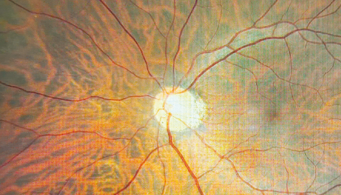
A recent observational study from Ophthalmology Retina has outlined a new morphologic staging system for full-thickness macular holes (FTMH) using spectral domain optical coherence tomography (SD-OCT) to track progressive outer retinal changes. The study analyzed SD-OCT scans of 52 patients over an extended period, defining four distinct stages of FTMH: Stage A shows initial separation of the retina from the retinal pigment epithelium (RPE) without retinal changes; Stage B involves thickening of the photoreceptor layer; Stage C displays patchy photoreceptor loss, creating a "moth-eaten" appearance; and Stage D represents extensive photoreceptor loss with a bare external limiting membrane (ELM).

The multi-institutional team of authors found that higher FTMH stages at diagnosis correlated with longer symptom duration and worse baseline visual acuity, which also predicted poorer visual outcomes after surgery. Patients at Stages B or C had better postoperative visual recovery compared to those in Stage D. Overall, surgery success rates were high, with 86.5 percent achieving anatomical closure, although delays in surgery were associated with advanced degeneration and worse recovery outcomes.

The authors believe that the staging system provides a reliable imaging biomarker for assessing FTMH progression, offering valuable guidance for surgical planning. Their work helps to emphasize the importance of early intervention, with the extent of photoreceptor damage observed impacting postoperative outcomes.

This SD-OCT-based classification also aligns with histological findings, potentially enabling clinicians to predict visual recovery more accurately and tailor treatment to individual patients.

About the Author(s)

The Ophthalmologist

More Articles by The Ophthalmologist

Related Content

Newsletters

Receive the latest Ophthalmology news, personalities, education, and career development – weekly to your inbox.

Newsletter Signup Image

False

Advertisement

False

Advertisement

Explore More in Ophthalmology

Dive deeper into the world of Ophthalmology. Explore the latest articles, case studies, expert insights, and groundbreaking research.

False

Advertisement
The Ophthalmologist
Subscribe

About

  • About Us
  • Work at Conexiant Europe
  • Terms and Conditions
  • Privacy Policy
  • Advertise With Us
  • Contact Us

Copyright © 2025 Texere Publishing Limited (trading as Conexiant), with registered number 08113419 whose registered office is at Booths No. 1, Booths Park, Chelford Road, Knutsford, England, WA16 8GS.

Disclaimer

The Ophthalmologist website is intended solely for the eyes of healthcare professionals. Please confirm below: