ANTERION from Heidelberg Engineering unites the power of high-resolution swept-source OCT images and precise measurements for the most important anterior segment examinations in one modular, upgradeable platform. Its recent software upgrade includes three new clinical features that further improve the quality of cornea and biometry data, and help avoid refractive surprises.
Uncover… the corneal epithelium
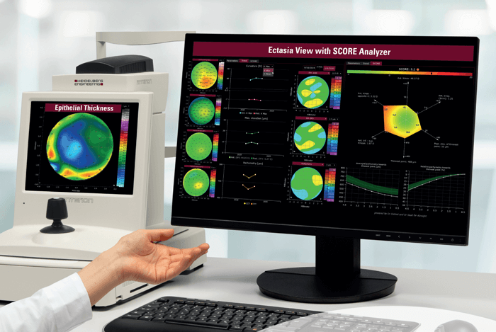
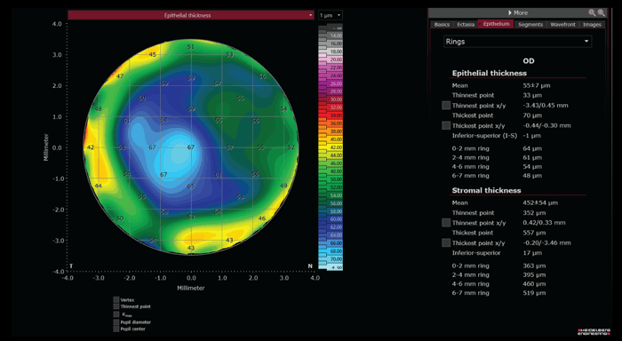
The additional Epithelial Thickness Module, with new parameters and color maps, facilitates thorough assessment of the epithelial thickness of a patient’s eyes. This feature can assist clinicians in refractive surgery planning and treatment control, ocular surface evaluation, ectasia screening, and other diagnostic areas. This new module for the ANTERION Cornea App also generates thickness maps and measurements for the residual stroma, providing further insights into patients’ corneal health.
Uncover… corneal ectasia
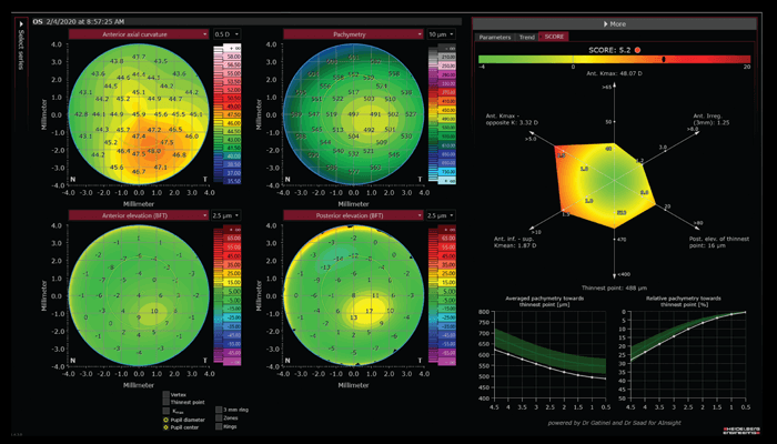
The new Ectasia View in ANTERION’s Cornea App aids in the assessment of ectatic changes in the cornea. The diagnostic dashboard combines all relevant information on one page. During a patient’s first visit, dedicated parameters and color maps for ectatic changes can be viewed and the values of both eyes can be compared. For follow-up visits, the dashboard offers intuitive visualizations of potential progression. The integrated SCORE Analyzer tool (Screening Corneal Objective Risk of Ectasia), developed by Damien Gatinel and Alain Saad, Paris, France, provides a unique means of evaluating keratoconus and other ectatic diseases. It combines multiple corneal indices that describe the magnitude of corneal steepening, thinning and asymmetry. An additional “radar map” as well as graphs for averaged and relative pachymetry provide a graphical presentation of relevant indices. Combined, the Ectasia View and integrated SCORE Analyzer provide modern aids in the assessment of ectatic diseases, such as forme fruste or advanced keratoconus, and help clinicians assess the probability of ectatic changes for their patients’ eyes.
Uncover….the scleral spur automatically
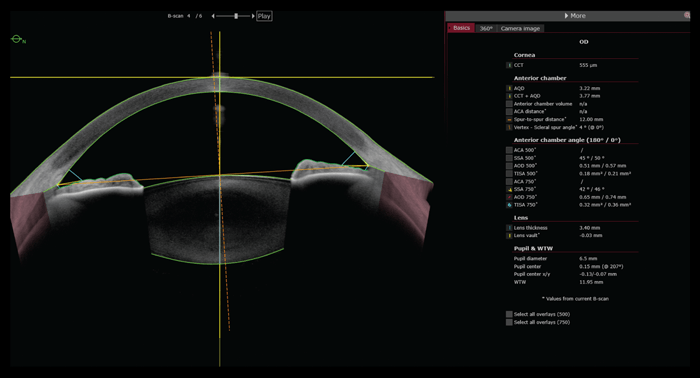
The powerful ANTERION Metrics App now also offers automatic detection of the scleral spurs after image acquisition. Capturing and detecting the precise position of this important landmark in the anterior chamber allows for automatic calculation and visualization of many biometric parameters, and thus facilitates clinical workflow. The manual resetting and modification of automatically set scleral spur markers remains possible.
Together, the impactful new features make ANTERION a truly complete anterior segment solution that reliably supports eye care professionals in multiple clinical disciplines in their delivery of patient care.
References
- Y Feng et al., “Heidelberg Anterion swept-source OCT corneal epithelial thickness mapping: repeatability and agreement with Optovue Avanti,” J Refract Surg, 38, 356 (2022). PMID: 35686707.
- R Herber et al., “Comparison of corneal tomography using a novel swept-source optical coherence tomographer and rotating Scheimpflug system in normal and keratoconus eyes: repeatability and agreement analysis,” Eye Vis (Lond), 9, 19 (2022). PMID: 35606839.
