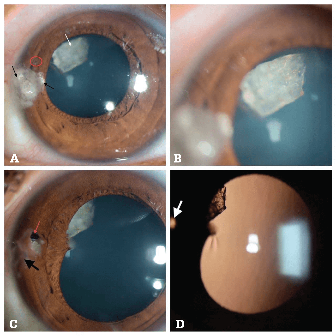

(A) Preoperative image of sealed corneal laceration with foreign body pieces and mucus embedded within the wound (thin black arrows); intralenticular foreign body (ILFB) seen out of focus (thin white arrow); though difficult to appreciate, a small iris hole is noted corresponding to the entry wound (within red circle).
(B) Magnified view showing the refractile nature of the ILFB.
(C) One month post-op, the healed wound is seen after suture removal (thick black arrow); iris defect is appreciated now (red arrow) with atrophy of iris tissue around it; the inert ILFB is partially obscured by focal posterior synechia. The remaining lens is clear, and there is no intraocular inflammation.
(D) Retroillumination view showing the ILFB partially obscured by posterior synechia and transillumination at the site of iris defect (thick white arrow).
Credit: Raksheeth Nathan Rajagopal and Muralidhar Ramappa, LV Prasad Eye Institute, Hyderabad, India.
References
- RN Rajagopal, M Ramappa, BMJ Case Rep, 14, e244875. PMID: 34479900.
