Conexiant
Login
  • Corneal Physician
  • Glaucoma Physician
  • New Retinal Physician
  • Ophthalmology Management
  • Ophthalmic Professional
  • Presbyopia Physician
  • Retinal Physician
The Ophthalmologist
  • Explore

    Explore

    • Latest
    • Insights
    • Case Studies
    • Opinion & Personal Narratives
    • Research & Innovations
    • Product Profiles

    Featured Topics

    • Anterior Segment
    • Glaucoma
    • Retina

    Issues

    • Latest Issue
    • Archive
  • Subspecialties
    • Cataract
    • Cornea
    • Glaucoma
    • Neuro-ophthalmology
    • Oculoplastics
    • Optometry
    • Pediatric
    • Retina
  • Business

    Business & Profession

    • Professional Development
    • Business and Entrepreneurship
    • Practice Management
    • Health Economics & Policy
  • Training & Education

    Career Development

    • Professional Development
    • Career Pathways

    Events

    • Webinars
    • Live Events
  • Events
    • Live Events
    • Webinars
  • Community

    People & Profiles

    • Power List
    • Voices in the Community
    • Authors & Contributors
  • Multimedia
    • Video
    • Podcasts
Subscribe
Subscribe

False

Advertisement
The Ophthalmologist / Issues / 2020 / Nov / Corneal Close-Up
Anterior Segment Cornea Business and Entrepreneurship Practice Management Sponsored

Corneal Close-Up

Navigate the cornea at a cellular level and select your preferred scanning depth for a comprehensive in vivo assessment of all layers – from epithelium to endothelium with the reintroduced HRT3 RCM

Sponsored By Heidelberg Engineering 11/12/2020 1 min read

Share

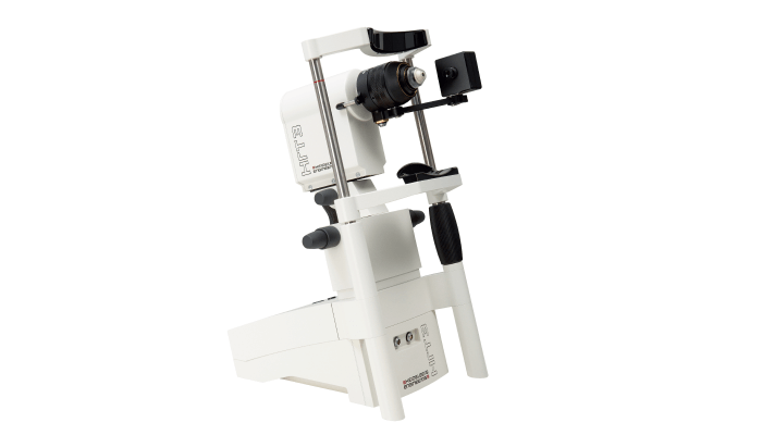
Reintroducing the HRT3 RCM by Heidelberg Engineering – a compact ophthalmic device that provides eye care specialists with a corneal microscope for the precise in vivo assessment of corneal layers and other ocular structures.

The HRT3 RCM uses confocal scanning laser microscopy to deliver high-resolution images of the cornea and external ocular structures, such as the conjunctiva or the limbus. Scanning the cornea with a field of view of up to 400 x 400 μm, the HRT3 RCM allows clinicians to acquire unique en face images of corneal cells, identify keratocytes subpopulations, and visualize details of the corneal subbasal nerve plexus.

Different acquisition modes further support the detailed analysis of cellular structures, allowing users to:
• Acquire single scans at any depth of the cornea
• Record a film sequence to dynamically explore areas of interest
• Perform a volume scan to receive a stack of images at different depths
• Use the semi-automated endothelial cell count to receive information about the morphology of this layer.

Comprehensive in vivo assessment with the HRT3 RCM empowers clinicians to diagnose and monitor corneal abnormalities, conduct pre- and post-surgery evaluations, analyze the corneal nerve structure in diabetic patients, and also evaluate dry eye disease.

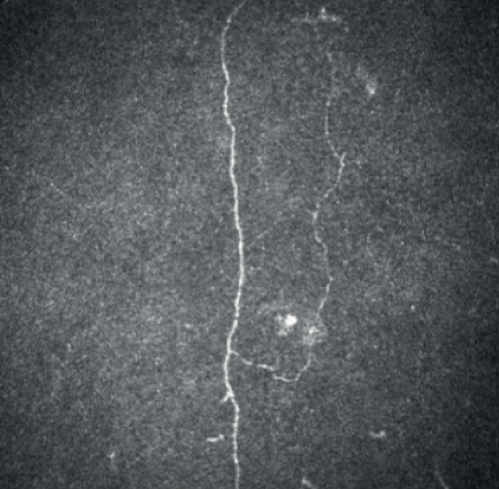
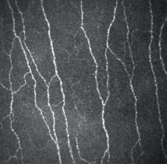
Assess corneal nerves at the microscopic level (Figures 1 and 2)

With the HRT3 RCM, it is possible to examine the corneal subbasal nerve plexus in detail; high-resolution images show the contrast in corneal nerve structures of a diabetic eye relative to a healthy eye.

Figure 1. Corneal nerve plexus in diabetic patient.
Figure 2. Corneal nerve plexus in healthy eye.

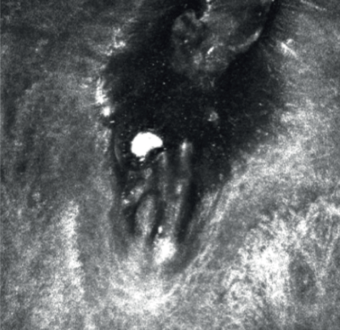
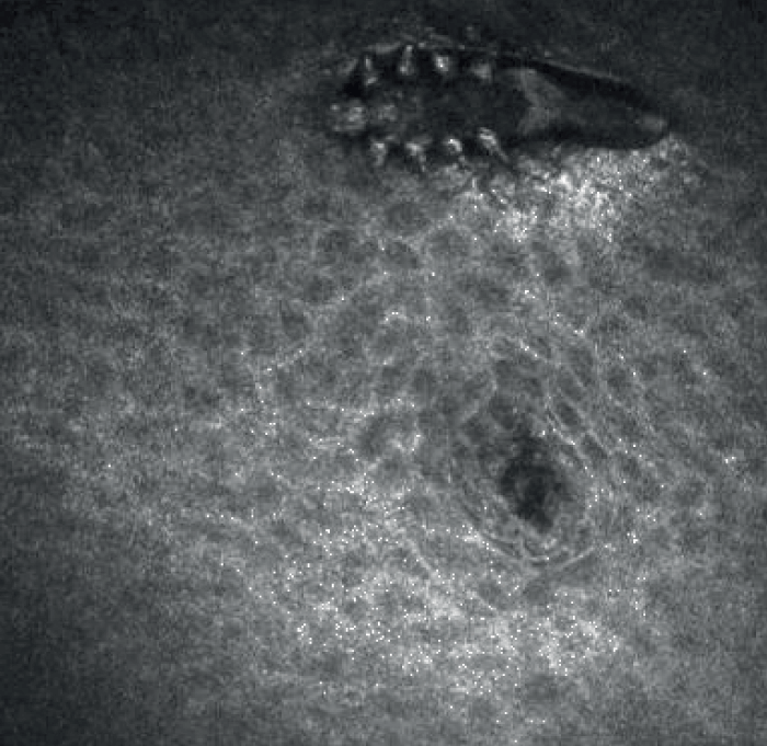
Investigate the conjunctiva, the limbus, the eye lid or meibomian glands to assess pathologies that affect these external ocular structures (Figures 3 and 4)

The HRT3 RCM can also aid in the assessment of Demodex blepharitis. These images show parts of a Demodex folliculorum and the whole body of a Demodex sebaceous that has climbed out of a meibomian gland orifice.

Figure 3. Demodex folliculorum.
Figure 4. Demodex sebaceous.

Related Content

Newsletters

Receive the latest Ophthalmology news, personalities, education, and career development – weekly to your inbox.

Newsletter Signup Image

False

Advertisement

False

Advertisement

Explore More in Ophthalmology

Dive deeper into the world of Ophthalmology. Explore the latest articles, case studies, expert insights, and groundbreaking research.

False

Advertisement
The Ophthalmologist
Subscribe

About

  • About Us
  • Work at Conexiant Europe
  • Terms and Conditions
  • Privacy Policy
  • Advertise With Us
  • Contact Us

Copyright © 2025 Texere Publishing Limited (trading as Conexiant), with registered number 08113419 whose registered office is at Booths No. 1, Booths Park, Chelford Road, Knutsford, England, WA16 8GS.

Disclaimer

The Ophthalmologist website is intended solely for the eyes of healthcare professionals. Please confirm below: