Conexiant
Login
  • Corneal Physician
  • Glaucoma Physician
  • New Retinal Physician
  • Ophthalmology Management
  • Ophthalmic Professional
  • Presbyopia Physician
  • Retinal Physician
The Ophthalmologist
  • Explore

    Explore

    • Latest
    • Insights
    • Case Studies
    • Opinion & Personal Narratives
    • Research & Innovations
    • Product Profiles

    Featured Topics

    • Anterior Segment
    • Glaucoma
    • Retina

    Issues

    • Latest Issue
    • Archive
  • Subspecialties
    • Cataract
    • Cornea
    • Glaucoma
    • Neuro-ophthalmology
    • Oculoplastics
    • Pediatric
    • Retina
  • Business

    Business & Profession

    • Professional Development
    • Business and Entrepreneurship
    • Practice Management
    • Health Economics & Policy
  • Training & Education

    Career Development

    • Professional Development
    • Career Pathways

    Events

    • Webinars
    • Live Events
  • Events
    • Live Events
    • Webinars
  • Community

    People & Profiles

    • Power List
    • Voices in the Community
    • Authors & Contributors
  • Multimedia
    • Video
    • Podcasts
Subscribe
Subscribe

False

Advertisement
The Ophthalmologist / Issues / 2020 / Jun / Early Days
Glaucoma

Early Days

The AI algorithm that can detect glaucomatous damage in retinal scans

By Aleksandra Jones 6/2/2020 1 min read

Share

Figure 1. A patient’s retina showing hyperfluorescent signals; each white spot is a single affected retinal nerve cell. Imagery supported by DARC technology, UCL/Western Eye Hospital.

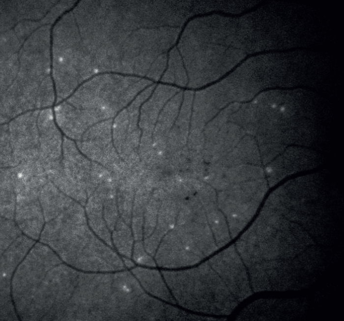
How useful would it be to have a test capable of picking up signs of glaucoma even 18 months earlier than with current methods, such as OCT? A new way of visualizing retinal cells, called DARC (Detection-of-Apoptosing-Retinal-Cells), developed as part of a clinical trial sponsored by the UCL Institute of Ophthalmology in London, UK, uses a fluorescent dye to illuminate cells in the process of apoptosis.

Then, an AI-aided algorithm analyzes patients’ retinal scans and refers those with higher DARC counts for further diagnosis. The trial found a significantly raised DARC number in patients who later progressed. The new method could also be used to test patients with AMD, as well as other neurodegenerative diseases.

References

  1. EM Normando et al., Expert Rev Mol Diagn, [Epub ahead of print] (2020). PMID: 32310684.

About the Author(s)

Aleksandra Jones

Having edited several technical publications over the last decade, I crossed paths with quite a few of Texere's current team members, and I only ever heard them sing the company's praises. When an opportunity arose to join Texere, I jumped at the chance! With a background in literature, I love the company's ethos of producing genuinely engaging content, and the fact that it is so well received by our readers makes it even more rewarding.

More Articles by Aleksandra Jones

Related Content

Newsletters

Receive the latest Ophthalmology news, personalities, education, and career development – weekly to your inbox.

Newsletter Signup Image

False

Advertisement

False

Advertisement

Explore More in Ophthalmology

Dive deeper into the world of Ophthalmology. Explore the latest articles, case studies, expert insights, and groundbreaking research.

False

Advertisement
The Ophthalmologist
Subscribe

About

  • About Us
  • Work at Conexiant Europe
  • Terms and Conditions
  • Privacy Policy
  • Advertise With Us
  • Contact Us

Copyright © 2025 Texere Publishing Limited (trading as Conexiant), with registered number 08113419 whose registered office is at Booths No. 1, Booths Park, Chelford Road, Knutsford, England, WA16 8GS.

Disclaimer

The Ophthalmologist website is intended solely for the eyes of healthcare professionals. Please confirm below: