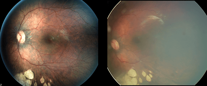
In 1998, Bert Massie PhD – the founder of Phoenix Technology Group – created the first digital camera to image the retinas of prematurely born babies, creating a new category in which digital images could be relied upon to help ophthalmic physicians screen for ROP and prevent blindness. In the years that followed, digital imaging replaced colored pencil drawings and became the standard of care for photo documentation and ROP screening. Dr. Massie left that business and formed Phoenix Technology Group in 2008. At Phoenix, he invented the first in vivo retinal imaging microscope to image the eyes of laboratory animals, transforming eye research and creating yet another new category. Today that product line – known as Phoenix MICRON – includes fundus imaging, FA, OCT and ERG. Knowing Dr. Massie’s reputation for innovation, a group of leading vitreoretinal surgeons got together in 2015 and suggested it was time for a new breakthrough in retinal imaging. Mobile phones had driven a revolution in digital imaging, and yet his 1998 invention had not changed. That was the genesis of the recently released, patented Phoenix ICON wide-angle retinal imaging camera. With the Phoenix ICON camera, Dr. Massie succeeded in delivering high-contrast, high-resolution retinal images, even on darkly pigmented retinas.
Dr. Massie approached the problem without the constraint of simply improving on an existing design. He completely reinvented the optics and camera system. Legacy systems inject light through the pupil at an angle, causing scatter as the returned light passes back to the camera system. To achieve high contrast, Dr. Massie and the team at Phoenix invented an optical system that uses annular illumination, establishing a clear, scatter-free return path “inside” the illumination ring. The team took the design even further by building a single-lens system with the magnification of a 30 degree lens, yet with a fully-illuminated 100 degree instantaneous field of view. This crucial step eliminated the onerous multi-lens process used in legacy cameras.
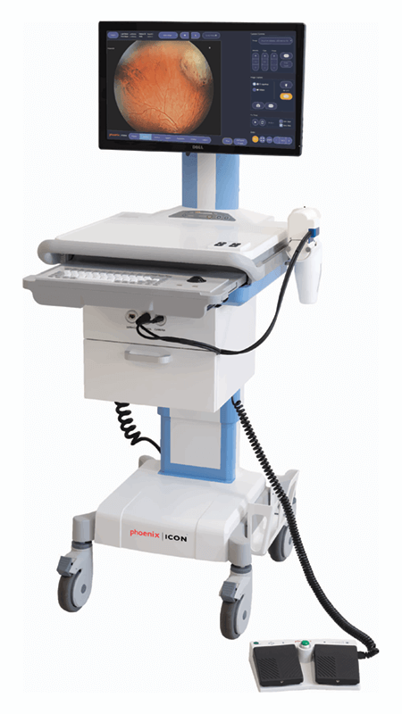
The Phoenix ICON delivers fundus imaging, and, by implementing interchangeable LED light modules, is capable of easily producing brilliant fluorescein angiograms. Simply change the light module in the hand piece, and flip the switch to position the barrier filter, and the operator is ready to capture angiograms.
The result? Stunning high-contrast, high-resolution fundus images and fluorescein angiograms, delivered from a single lens system capable of imaging for 6 hours on battery.
The Phoenix team was not finished innovating. The team recognized that the installed legacy imaging platforms are “islands” in the context of hospital and clinic information systems. Put another way, images were captured and stored on a local camera hard drive. Although they support “DICOM format,” the images needed to be manually exported to a thumb drive, and then manually uploaded to the hospital information system. No hospital IT person is happy with images moving around on a thumb drive – and clinicians are frustrated by the upload time.
Any change to image sharing processes need to take into consideration privacy, security and image management requirements. As a result, Phoenix has just announced a new DICOM connector for the Phoenix ICON. The DICOM connector completes the integration loop by implementing the DICOM networking protocol.
Now, with the new DICOM connector for the Phoenix ICON, an operator can select images from a study, push a button, and deliver those images to the hospital or clinic Photo Archive and Communications System (PACS). When images are in the PACS they can be accessed by all the constituents that need them for interpretation, documentation, and reporting. And that means the ICON camera saves time, eases the workflow, and complies with security, data retention, and patient information management required of hospital and clinic operations. The Phoenix team takes pride in being an innovator yet recognizes that without adoption, all the innovation in the world is meaningless. After over 20 years of innovating in the wide-angle, contact retinal imaging arena, Phoenix has earned the endorsements and accolades from a host of trailblazing ophthalmologists, including Dr. Mike Trese and Dr. Carol Shields, who use Phoenix ICON cameras to image their patients.