Conexiant
Login
  • Corneal Physician
  • Glaucoma Physician
  • New Retinal Physician
  • Ophthalmology Management
  • Ophthalmic Professional
  • Presbyopia Physician
  • Retinal Physician
The Ophthalmologist
  • Explore

    Explore

    • Latest
    • Insights
    • Case Studies
    • Opinion & Personal Narratives
    • Research & Innovations
    • Product Profiles

    Featured Topics

    • Anterior Segment
    • Glaucoma
    • Retina

    Issues

    • Latest Issue
    • Archive
  • Subspecialties
    • Cataract
    • Cornea
    • Glaucoma
    • Neuro-ophthalmology
    • Oculoplastics
    • Optometry
    • Pediatric
    • Retina
  • Business

    Business & Profession

    • Professional Development
    • Business and Entrepreneurship
    • Practice Management
    • Health Economics & Policy
  • Training & Education

    Career Development

    • Professional Development
    • Career Pathways

    Events

    • Webinars
    • Live Events
  • Events
    • Live Events
    • Webinars
  • Community

    People & Profiles

    • Power List
    • Voices in the Community
    • Authors & Contributors
  • Multimedia
    • Video
    • Podcasts
Subscribe
Subscribe

False

Advertisement
The Ophthalmologist / Issues / 2018 / May / DIY OCT
Retina

DIY OCT

A hand-held device that aims to reduce the disease burden for AMD patients

By Ruth Steer 5/23/2018 1 min read

Share

Figure 1. a. The current prototype for the hand-held device. Credit: Universitätsklinikum Schleswig-Holstein, Kiel, and Medical Laser Center, Lübeck, Germany.

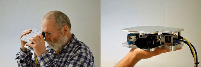
AMD may well be treatable with anti-VEGF injections, but it comes at a cost to the patient: regular visits to the ophthalmologist’s office for disease monitoring with OCT. For elderly and visually impaired patients who require travel assistance, these frequent appointments can represent a further burden on their relatives – or their finances. A team from Universitätsklinikum Schleswig-Holstein, Kiel, and Medical Laser Center, Lübeck, Germany, have a potential solution in the form of a hand-held OCT device for home-monitoring of patients with retinal disease. Given today’s advanced – and costly – OCT systems, what does a home-care setup look like, and how can it be achieved? “For a device to be used by the patients themselves, it needs to be small and low-cost,” explains Claus von der Burchard, Research Fellow at Universitätsklinikum Schleswig-Holstein. “To get there, we’ve had to make some compromises on image quality.” The result is off-axis full-field time-domain OCT (1). Von der Burchard explains: “We focused on reducing the scan area to 3 x 3 mm and using a full-field system which illuminates the whole field.” According to von der Burchard, the 3 x 3 images provide good sensitivity and specificity for monitoring subretinal and intraretinal fluid volume, and the full-field approach increases visual quality. “The design is also very simple, needing only a standard light source – rather than swept-source – and a regular, low-cost USB camera.”

Figure 1. b. SD-OCT image (left) and off-axis full-field time domain OCT image (right) from a patient with AMD (84 years old; visual acuity, 20/80). The red arrow depicts subretinal fluid which is visible on images from both the clinical and hand-held device.

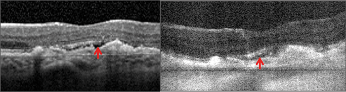
Though the team are still in the research phase, they have tested their device – alongside spectral-domain (SD)-OCT – in 10 patients with retinal disease (including AMD and retinal vein occlusion), and have shown that it can acquire clinically-useful images (Figure 1). “Even though the image quality is not quite as good as clinical OCT systems, the subretinal fluid does demark very well and the need for re-treatment is apparent,” says von der Burchard. The team plans to continue developing their device, and study its imaging capabilities in more patients by asking clinicians to grade both full-field OCT and SD-OCT images (while being masked), and compare their image quality, which biomarkers are present, and  whether the patient needs anti-VEGF retreatment. “We believe that a hand-held OCT device would have a huge impact on alleviate disease burden, and may represent a paradigm shift in the treatment of wet AMD and other diseases.”

References

  1. H Sudkamp et al., “In-vivo retinal imaging with off-axis full-field time-domain optical coherence tomography”, Opt Lett, 41, 4987–4990. PMID: 27805666.

About the Author(s)

Ruth Steer

More Articles by Ruth Steer

Related Content

Newsletters

Receive the latest Ophthalmology news, personalities, education, and career development – weekly to your inbox.

Newsletter Signup Image

False

Advertisement

False

Advertisement

Explore More in Ophthalmology

Dive deeper into the world of Ophthalmology. Explore the latest articles, case studies, expert insights, and groundbreaking research.

False

Advertisement
The Ophthalmologist
Subscribe

About

  • About Us
  • Work at Conexiant Europe
  • Terms and Conditions
  • Privacy Policy
  • Advertise With Us
  • Contact Us

Copyright © 2025 Texere Publishing Limited (trading as Conexiant), with registered number 08113419 whose registered office is at Booths No. 1, Booths Park, Chelford Road, Knutsford, England, WA16 8GS.

Disclaimer

The Ophthalmologist website is intended solely for the eyes of healthcare professionals. Please confirm below: