When it comes to retinal disease, information is key. Knowing more about the health of the vision system enables early detection, as well as enhanced patient management post-treatment, which leads to better visual outcomes for the patient. As the world-leader in modern visual electrophysiology, Diopsys®, Inc., has done more than any other company to advance the use of electroretinography (ERG) and visual evoked potential (VEP) in eye care practices – and provide eye care professionals with objective and functional information on the health of their patients’ vision systems.
With OCT providing objective structural information, fundus photography providing subjective structural information, and visual field testing providing subjective functional information, modern electrophysiology provides the objective functional information that eye care providers need – filling the ‘information void’ in most practices. Diopsys’ unique patented technology was created with practice-based eye care professionals in mind, and features space-efficient designs, streamlined testing protocols, and intuitive color-coded reports based on documented reference ranges. Three unique platforms – Diopsys® NOVA™ (cart), Diopsys® ARGOS™ (tabletop) and the NEW Diopsys® RETINA PLUS™ (carry case) – accommodate practices of all sizes, and each system can be customized with a full suite of visual electrophysiology modules:
- Diopsys® ffERG / Flicker (full field ERG) tests record the retina’s response to flashes of light – reflecting the electrical activity of cone and bipolar cells – and provide objective functional information about global retinal health. Clinically effective in helping to manage retinal disorders, such as diabetic retinopathy, central retinal vein occlusion, retinal concerns obscured by media opacities and uveitis (1)(2)(3)(4)(5), Flicker vision tests use intuitive color-coded reports to help physicians evaluate retinal disease severity (1)(2), predict retinal ischemia (2)(3)(4), quantify retinal function loss and recovery (1)(2)(5), and monitor retinal function for appropriate and timely treatment (1)(2)(5).
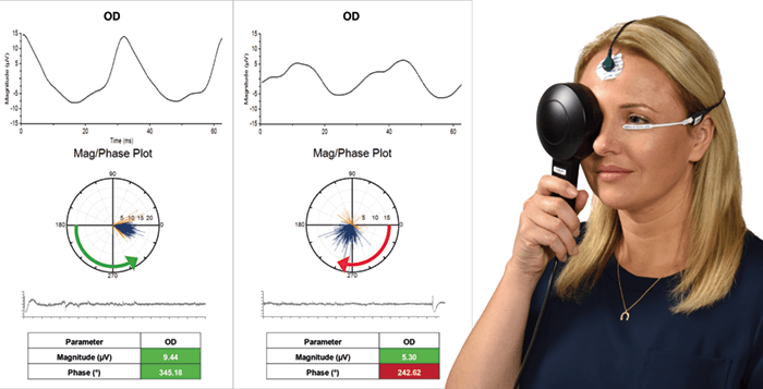
- Diopsys® ERG (pattern ERG) tests record the retina’s response to a phase-reversing patterned stimulus and provide objective functional information on the performance of retinal ganglion cells. Effective in helping physicians diagnose and manage diseases including glaucoma, age-related macular degeneration (AMD) and diabetic edema (6)(7)(8), ERG tests identify ‘stressed’ cells at a subclinical stage – sometimes years earlier than OCT (6). Starting treatment at this early stage – before evident structural defects – can help extend patient sight.
- Diopsys® mfERG (multifocal ERG) provides objective information on localized retinal function, and helps physicians recognize the first signs of drug-induced retinopathy (9)(10). As most ocular side effects of drug-induced toxicity are reversible after cessation of therapy if detected early (10), mfERG can help physicians co-manage patients more efficiently.
- Diopsys® VEP (visual evoked potential) testing objectively records the conductivity of the visual pathway in response to a phase-reversing patterned stimulus, and provides important diagnostic information on the functional integrity of the entire visual system, from the anterior segment to the visual cortex. Useful for detecting and monitoring many types of visual abnormalities, it is often used for the diagnosis and management of neuro-visual disorders, such as optic neuritis, amblyopia, and vision problems caused by traumatic brain injury (11)(12)(13).
Testimonials
“The only objective, functional method at our disposal for diagnosing and managing retinopathies and glaucoma is ERG. Thankfully, scientific advances from Diopsys® have enabled this testing modality to be incorporated into the clinic. ERG testing has more than proven its worth in evaluating retinal pathologies, which means that I use the Diopsys® device in my clinic daily.” William Ayliffe, FRCS, PhD, Consultant Ophthalmologist, Lister Hospital, London, UK. “Accessible, modern visual electrophysiology devices from Diopsys® provide objective measures of function and are useful in following progression of disease, or response to treatment. Pattern ERG is particularly useful when visual field testing is difficult to perform. Most importantly, though, it provides a mechanism to diagnose glaucoma at an earlier stage – when it is in a reversible state. Thus, pattern ERG testing represents a powerful tool to aid clinical decision making, especially regarding when to start therapy to avoid vision loss from glaucoma progression.” André Mermoud, MD, Medical Director, Genolier Swiss Visio Network SA, Clinique de Montchoisi, Lausanne, Switzerland.www.diopsys.com
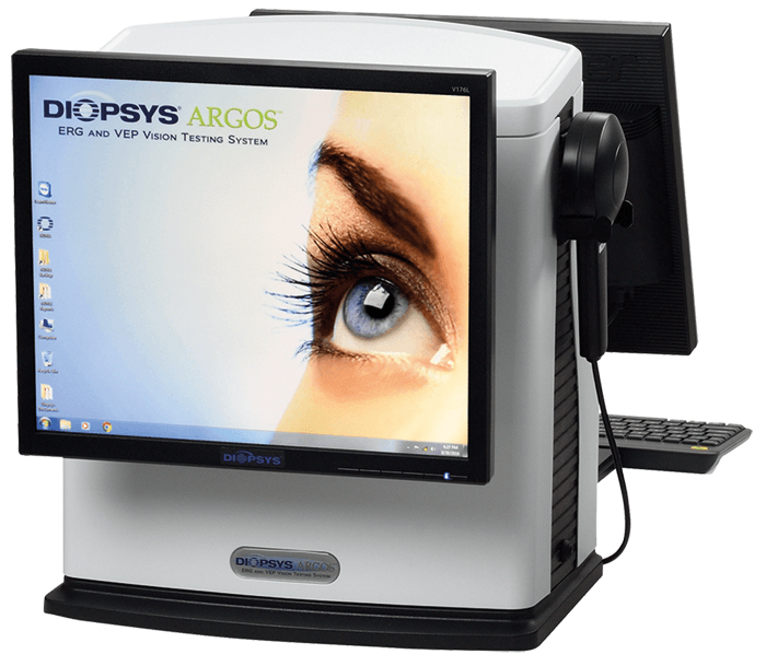
The Diopsys® NOVA is an electrophysiology device that generates photic stimuli, and records, processes, and analyzes the resultant signals to provide information about the visual system. Availability varies globally.References
- S Yasuda et al., Acta Ophthalmol, e465–468 (2015). PMID: 25597703. MM Moschos et al., Clin Ophthalmol, 8, 199–214 (2014). PMID: 24453476. J Larsson and S Andreasson. Br J Ophthalmol, 85, 683–685 (2001). PMID: 11371488. T Ratanapakorn et al., J Med Assoc Thai, 93, 1196–1199 (2010). PMID: 20973323. K Holm et al., DOC Ophthalmol, 131, 43–451 (2015). PMID: 25773362. MR Banitt et al, IOVS, 54, 2346–2352 (2013). PMID: 23412088. A Oner et al. Doc Ophthalmol, 119, 37–42 (2009). PMID: 19225818. A Ozkiris. Doc Ophthalmol, 120, 243–250 (2010). PMID: 20140695. DC Hood et al., Doc Ophthalmol, 124, 1–13 (2012). PMID: 22038576. M Dettoraki and MM Moschos. Ophthalmic Res, 56, 169–177 (2016). PMID: 27351191. RT Naismith et al., Neurology, 73, 46–52 (2009). PMID: 19564583. JW Simon et al., J AAPOS, 8, 549–554 (2004). PMID: 15616502. M McKerral et al., IOVS,43, e-abstract 1803 (2002).
