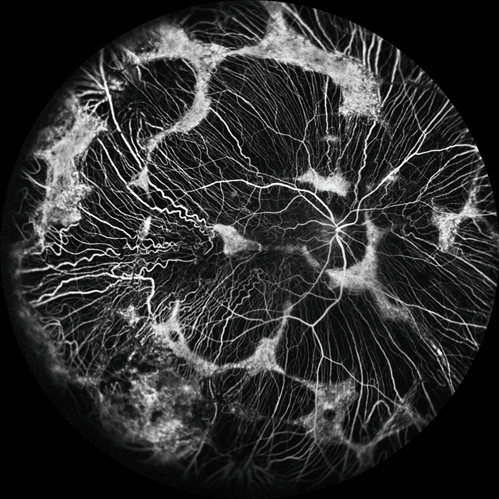
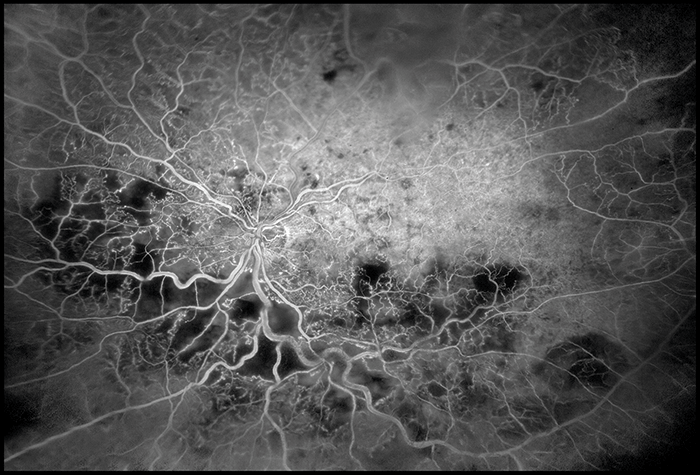
Choroideremia “Anticipation and timing is key in my work as both a professional sports photographer and as an ophthalmic photographer. Anticipating and capturing the dynamic flow of fluorescein through retinal blood vessels is important when performing angiography. In this case of choroideremia, both the retinal and choroidal vessels are visible along with arc-like remnants of what remains of the RPE.” Joe Territo, senior photographer, Retina Associates of Western New York, Rochester, NY, USA. His personal work can be viewed at: http://joeterritophotography.com/
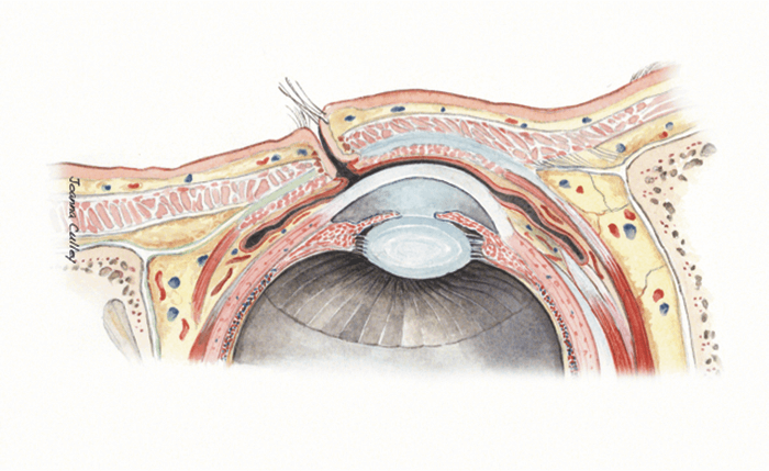
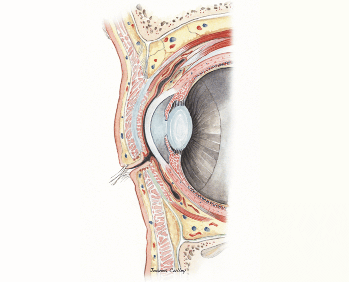
Anatomy Artistry Watercolour illustration by Medical Artist Joanna Culley of www.medical-artist.com. Joanna Culley, Medical Artist, Haslemere, Surrey, UK.
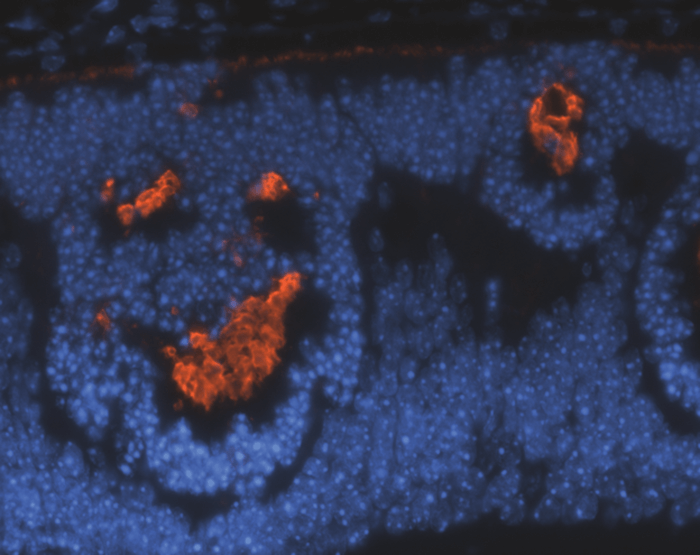
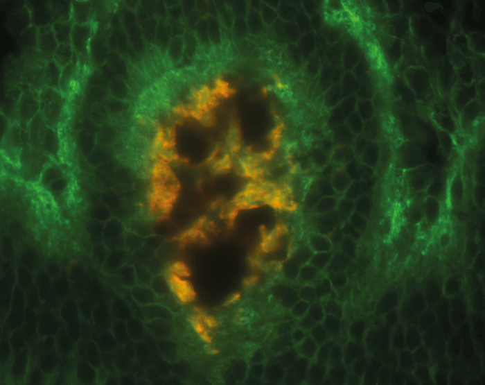
Seeing Faces Histological retinal sections from mice that have had NRL and RDS proteins knocked out that appear to depict, respectively, a “happy” and a “monkey/ghost” face (images above). The top image shows photoreceptor outer segments labeled with anti-S-opsin antibody (red) and the bottom image shows photoreceptor inner segments labeled with anti Na+/K+-ATPase antibody (green) and outer segments labelled with anti-S-opsin (red). Rafal Farjo, CEO of EyeCRO LLC and Charlesson LLC, Oklahoma City, OK, USA.
This selection of images was contributed by Kim Baxter, Ophthalmic Photography Team Leader at Addenbrooke’s Hopsital, Cambridge, UK. Baxter, who specializes in all types of ophthalmic imaging including slit-lamp photography and retinal angiography, is a member of the Institute of Medical Illustrators and has won two gold awards at their annual exhibitions, along with bronze and silver awards. Her work has been selected for the Wellcome Trust and twice by the Royal Photographic Society’s Visualising Medicine exhibitions. Kim Baxter, Ophthalmic Photography Team Leader at Addenbrooke’s Hospital, Cambridge, UK.
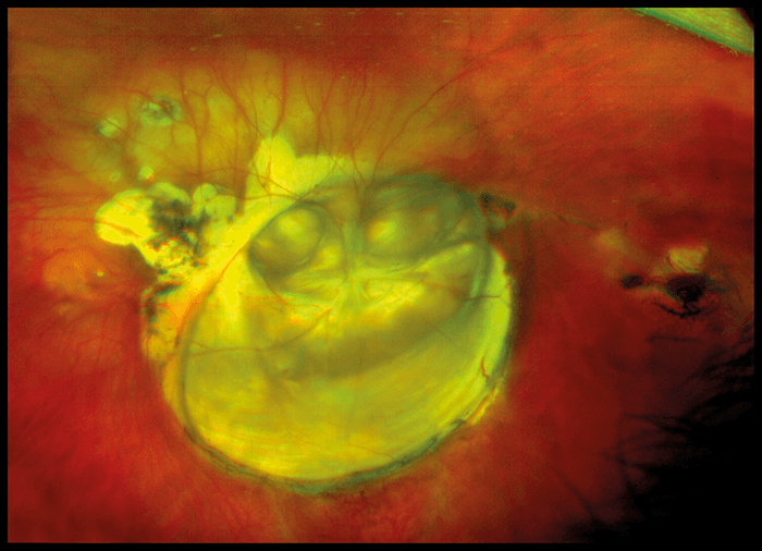
Coloboma Close-up This coloboma covers the macula area, including the optic nerve, and was captured with ultra-widefield imaging.
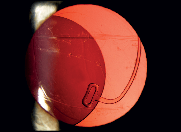
Subluxed IOL This image of a subluxed IOL was taken on an anterior segment camera using retroillumination to show the position of the lens prior to surgery.
Seeing CRVO Ischemic central retinal vein occlusion taken on a wide-field camera during a fluorescein angiogram to document the level of ischemia before treatment.
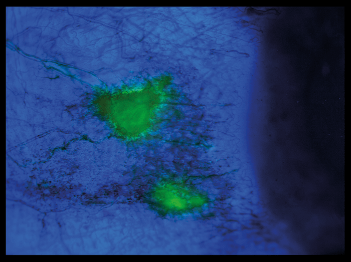
Imaging Infection Using an anterior segment camera, topical fluorescein dye and blue filters, the areas of conjunctival infection can be seen in green.