Conexiant
Login
  • Corneal Physician
  • Glaucoma Physician
  • New Retinal Physician
  • Ophthalmology Management
  • Ophthalmic Professional
  • Presbyopia Physician
  • Retinal Physician
The Ophthalmologist
  • Explore

    Explore

    • Latest
    • Insights
    • Case Studies
    • Opinion & Personal Narratives
    • Research & Innovations
    • Product Profiles

    Featured Topics

    • Anterior Segment
    • Glaucoma
    • Retina

    Issues

    • Latest Issue
    • Archive
  • Subspecialties
    • Cataract
    • Cornea
    • Glaucoma
    • Neuro-ophthalmology
    • Oculoplastics
    • Pediatric
    • Retina
  • Business

    Business & Profession

    • Professional Development
    • Business and Entrepreneurship
    • Practice Management
    • Health Economics & Policy
  • Training & Education

    Career Development

    • Professional Development
    • Career Pathways

    Events

    • Webinars
    • Live Events
  • Events
    • Live Events
    • Webinars
  • Community

    People & Profiles

    • Power List
    • Voices in the Community
    • Authors & Contributors
  • Multimedia
    • Video
    • Podcasts
Subscribe
Subscribe

False

Advertisement
The Ophthalmologist / Issues / 2017 / Jan / The Clear Lens Warden
Anterior Segment Cataract Research & Innovations

The Clear Lens Warden

A gene associated with early-onset Parkinson’s disease appears to act against cataract formation

By Roisin McGuigan 1/11/2017 1 min read

Share

Figure 1. When LECs expressing wild-type parkin protein are exposed to hydrogen peroxide-induced oxidative stress, parkin colocalizes with mitochondria and recruits proteins to degrade the damaged mitochondria (yellow puncta) (a) while LECs expressing dysfunctional mutated parkin (C431N) do not (b). (Credit: Florida Atlantic University.)

Parkin is an interesting protein. Encoded in humans by the PARK2 gene, it is implicated in several disease states, including Parkinson’s disease. How PARK2 mutations lead to dopaminergic cell death and early Parkinsonian symptoms isn’t clear, though – it appears to play a role in the degradation of free-radical-damaged mitochondria, but what’s now clear is that parkin also plays a central role in keeping the lens… clear.

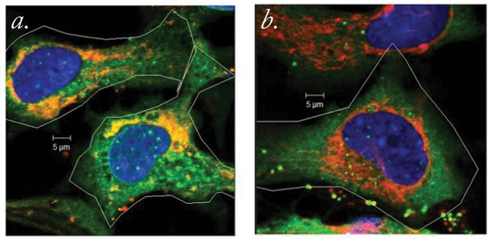
Intrigued by the protein’s potential role in lens opacity, researchers from the Charles E Schmidt College of Medicine at Florida Atlantic University decided to delve deeper. They performed cell culture experiments in which lens epithelial cells (LECs) expressing either normal or mutated forms of the PARK2 gene were assessed. What they found was this: PARK2 is expressed when LECs are exposed to cataract-causing, free radical-generating environmental insults (in this case, oxidative stress caused by hydrogen peroxide exposure). Parkin removes damaged mitochondria (see Figure 1), and by doing so, helps prevent the formation of free radicals in LECs. This increases the ability of the LECs to survive free radical formation, and presumably, the reactive oxygen species-mediated aging.

“Our findings suggest that parkin plays a direct role in the prevention of oxidative stress through its ability to maintain cellular mitochondrial populations, and that the gene encoding parkin is induced by environmental damage,” says Marc Kantorow, lead author of the associated paper (1). What could this mean? “Drugs or genetic methods that increase parkin levels and function could prove effective in preventing cataracts and other age-related degenerative diseases, including neurological conditions like Parkinson’s disease,” he explains. According to Kantorow, the team now plans to “establish how parkin regulates the growth and development of the lens by controlling mitochondrial populations that are required for lens cell growth.” He adds, “We want to identify the genetic mechanisms that regulate the production of parkin in cells and see if they can be manipulated to increase parkin levels, thereby increasing cell survival to prevent disease.”

References

  1. L Brennan et al., “Parkin elimination of mitochondria is important for maintenance of lens epithelial cell ROS levels and survival upon oxidative stress exposure”, Biochim Biophys Acta, 1863, 21–32 (2017). PMID: 27702626.

About the Author(s)

Roisin McGuigan

I have an extensive academic background in the life sciences, having studied forensic biology and human medical genetics in my time at Strathclyde and Glasgow Universities. My research, data presentation and bioinformatics skills plus my ‘wet lab’ experience have been a superb grounding for my role as a deputy editor at Texere Publishing. The job allows me to utilize my hard-learned academic skills and experience in my current position within an exciting and contemporary publishing company.

More Articles by Roisin McGuigan

Related Content

Newsletters

Receive the latest Ophthalmology news, personalities, education, and career development – weekly to your inbox.

Newsletter Signup Image

False

Advertisement

False

Advertisement

Explore More in Ophthalmology

Dive deeper into the world of Ophthalmology. Explore the latest articles, case studies, expert insights, and groundbreaking research.

False

Advertisement
The Ophthalmologist
Subscribe

About

  • About Us
  • Work at Conexiant Europe
  • Terms and Conditions
  • Privacy Policy
  • Advertise With Us
  • Contact Us

Copyright © 2025 Texere Publishing Limited (trading as Conexiant), with registered number 08113419 whose registered office is at Booths No. 1, Booths Park, Chelford Road, Knutsford, England, WA16 8GS.

Disclaimer

The Ophthalmologist website is intended solely for the eyes of healthcare professionals. Please confirm below: