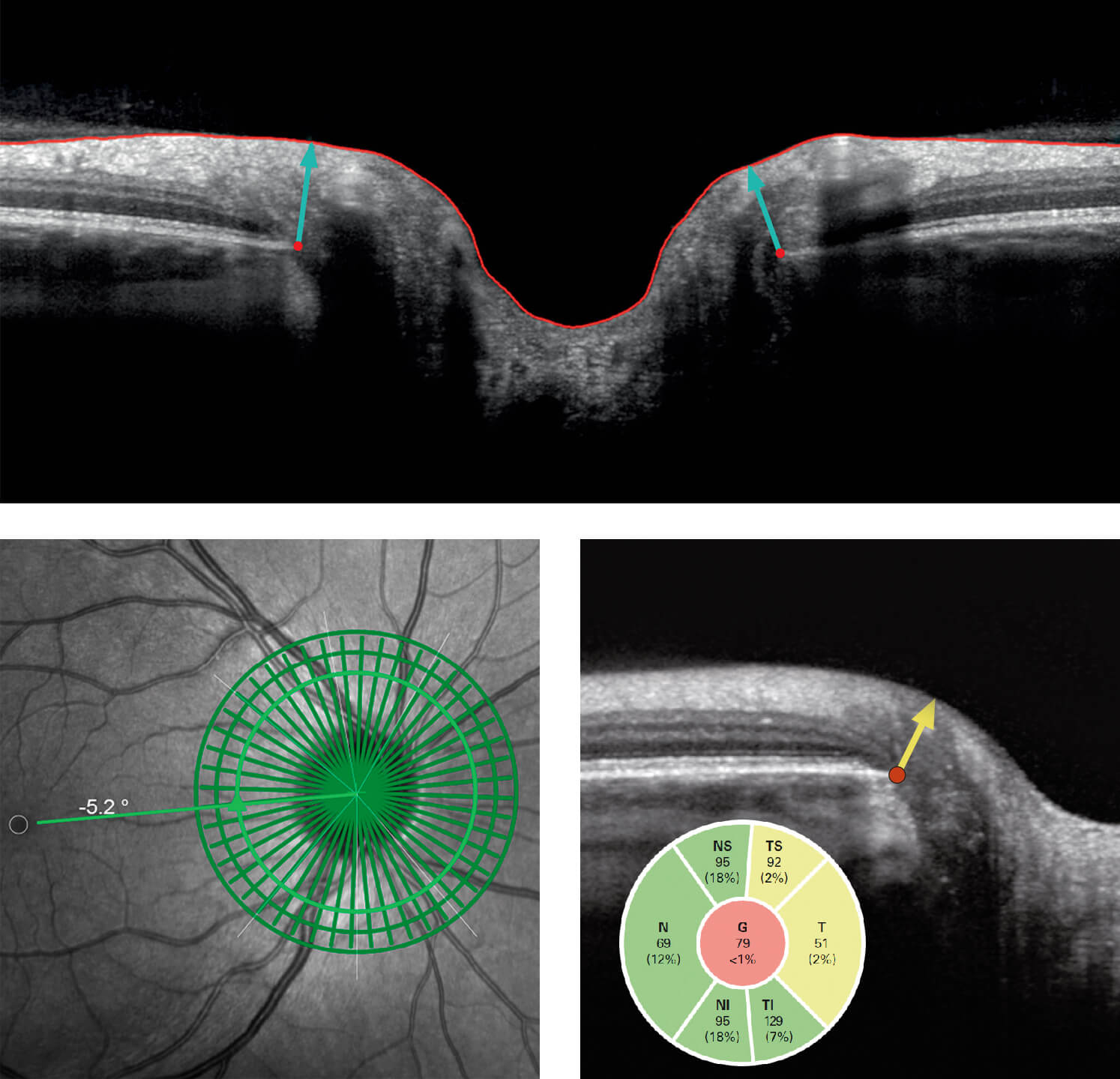
The Glaucoma Module Premium Edition for SPECTRALIS provides a comprehensive analysis of the optic nerve head, retinal nerve fiber layer, and ganglion cell layer by precisely matching unique scan patterns to the fine anatomic structures relevant in glaucoma diagnostics. By means of a new proprietary technology called the Anatomic Positioning System, the module creates an anatomic map unique to each patient’s eye using two fixed, structural landmarks: the center of the fovea and the center of Bruch’s membrane opening. All subsequent scan protocols are automatically oriented according to each eye’s unique anatomic map, enabling a precise examination of relevant structures over time. Furthermore, the Glaucoma Module Premium Edition compares patients’ eyes to an age- and disc size-adjusted reference database of healthy eyes representing the racial and ethnic mix of the US population, flagging small deviations that may be clinically relevant. This allows for a highly sensitive assessment of structural damage characteristic of glaucoma and a close monitoring of structural changes indicative of glaucomatous progression. The physiological and technical premise of the Glaucoma Module Premium Edition is based on research collaborations between two groups led by Balwantray C. Chauhan, Ph.D., Dalhousie University in Halifax (Canada), and Claude F. Burgoyne, M.D., Devers Eye Institute in Portland, OR (USA) performed with the SPECTRALIS imaging platform1,2,3,4,5,6. Their work revealed the importance of making an anatomically and geometrically accurate neuroretinal rim measurement using Bruch’s membrane opening, the anatomical outer border of the rim. “The measurements are more accurate because they are based on an identifiable anatomical border of the rim and take into account its varying geometry at the point of measurement”, said Dr. Burgoyne. Dr. Chauhan added: “We have already been able to demonstrate that the use of this new measurement method translates into significantly enhanced diagnostic accuracy.” “The 510(k) clearance of this product is great news for our company and also for glaucoma patients in the U.S. The Glaucoma Module Premium Edition is an excellent tool to assess glaucomatous damage based on a novel objective examination of the optic nerve head anatomy”, summarizes Dr. Gerhard Zinser, Managing Director of Heidelberg Engineering. References:
- Chauhan B.C. et al., Ophthalmology, 2013;120:535–543.
- Chauhan B.C. and Burgoyne C.F., Am J Ophthalmol. 2013;156:218-227.
- Chauhan B.C. et al.,Ophthalmology, 2015;122:1786-1794.
- He L. et al., PLoS One, 2014;9:e92225.
- Reis A.S.et al., Ophthalmology, 2012;119:738–747.
- Reis A.S. et al., Invest. Ophthalmol. Vis. Sci., 2012;53:1852–1860.
Heidelberg Engineering www.HeidelbergEngineering.com
