Conexiant
Login
  • Corneal Physician
  • Glaucoma Physician
  • New Retinal Physician
  • Ophthalmology Management
  • Ophthalmic Professional
  • Presbyopia Physician
  • Retinal Physician
The Ophthalmologist
  • Explore

    Explore

    • Latest
    • Insights
    • Case Studies
    • Opinion & Personal Narratives
    • Research & Innovations
    • Product Profiles

    Featured Topics

    • Anterior Segment
    • Glaucoma
    • Retina

    Issues

    • Latest Issue
    • Archive
  • Subspecialties
    • Cataract
    • Cornea
    • Glaucoma
    • Neuro-ophthalmology
    • Oculoplastics
    • Optometry
    • Pediatric
    • Retina
  • Business

    Business & Profession

    • Professional Development
    • Business and Entrepreneurship
    • Practice Management
    • Health Economics & Policy
  • Training & Education

    Career Development

    • Professional Development
    • Career Pathways

    Events

    • Webinars
    • Live Events
  • Events
    • Live Events
    • Webinars
  • Community

    People & Profiles

    • Power List
    • Voices in the Community
    • Authors & Contributors
  • Multimedia
    • Video
    • Podcasts
Subscribe
Subscribe

False

Advertisement
The Ophthalmologist / Issues / 2016 / Jun / The Diagnostic Dome

The Diagnostic Dome

A new device could overcome the challenges of performing perimetry in infants

By Mark Hillen 6/9/2016 1 min read

Share

Figure 1. Pediatric Perimeter device showing the hemispherical dome and infrared camera placement. The LEDs are arranged to allow visual field measurement at 10° radial intervals; upper, left and right visual fields are measurable out to 80° and lower visual field measurable to 50°.

Visual field assessment in infants is hard… and perimetry is incredibly difficult (1). But it’s important information to have when visual field disorders or strabismus is suspected, and it can help identify and exclude those cases where infants present with an “eye turn”, receive strabismus surgery, but actually have congenital hemianopia.

The Pediatric Perimeter’s software in use during visual field mapping.

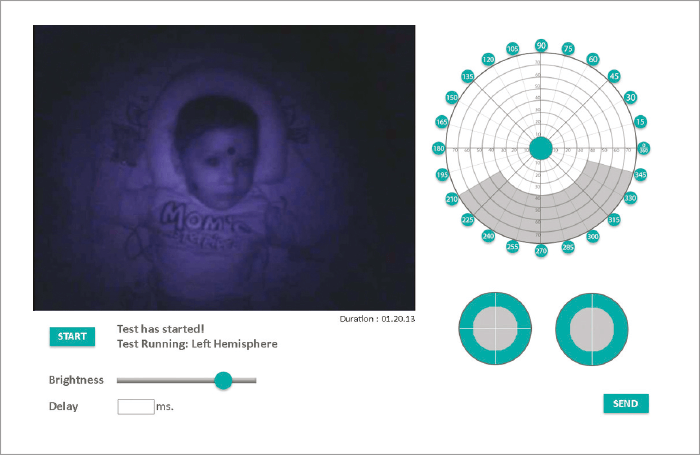
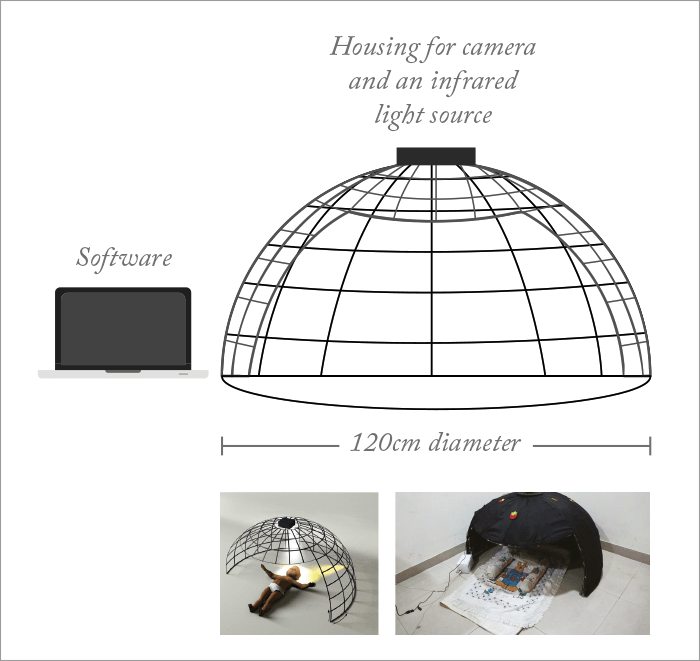
What if there was an easier way? The Pediatric Perimeter (1) is a hemispherical dome built out of steel rods to form 24 meridians (separated by 15°), that has an infrared (IR) camera at its apex, and is covered by black cloth (Figure 1). The infant is laid in the dome, and the infrared camera monitors their eye movements in response to LED stimulus patterns controlled by the clinician (Figure 1). Does it work? PremNandhini Satgunam of the L V Prasad Eye Institute in Hyderabad, India, and a lead member of the development team, explained that the perimeter has been used to successfully map visual field extent in both infants and toddlers. Further, it’s able to identify differences in the visual fields of infants with normal developmental milestones (NM) and those with developmental delay (DD). Five NM infants (mean age, 10.0±7.1 months) and 12 DD infants (mean age, 12.1±6.9 months) were assessed.

Figure 2. Median reaction time of infants with normal milestones and developmental delay measured during pediatric perimetry. DD, developmental delay; NM, normal milestones.

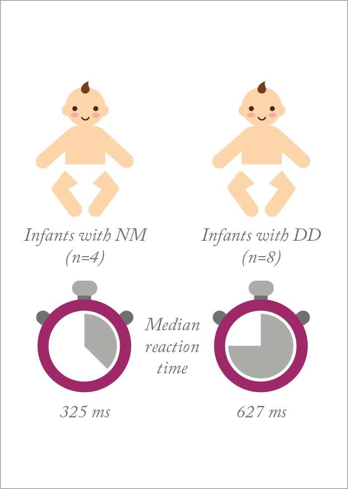
Although both groups displayed similar visual field extents, reaction times in DD infants were almost two times longer (Figure 2) – something that Satgunam ascribed to “potential processing delays in afferent, efferent, or both systems.” The team also showed preliminary findings that the visual field isopter may differ between infants with NM and DD, but Satgunam comments, “we need to do a lot more normative database measurement before we can say anything for certain.” If pediatric visual development is better understood, in both normal and DD children, as well as furthering knowledge to guide clinical practice, it may prevent unnecessary surgeries being performed on young children. 

References

  1. V Dobson et al., “Visual field extent in children 3.5–30 months of age tested with a double-arc LED perimeter”, Vis Res, 38, 2743–2760 (1998). PMID: 9775323. P Satgunam et al., “Development of a novel device to measure visual fields in infants and toddlers”, IOVS 2016, 57, ARVO E-Abstract 5978-D0069.

About the Author(s)

Mark Hillen

I spent seven years as a medical writer, writing primary and review manuscripts, congress presentations and marketing materials for numerous – and mostly German – pharmaceutical companies. Prior to my adventures in medical communications, I was a Wellcome Trust PhD student at the University of Edinburgh.

More Articles by Mark Hillen

Related Content

Newsletters

Receive the latest Ophthalmology news, personalities, education, and career development – weekly to your inbox.

Newsletter Signup Image

False

Advertisement

False

Advertisement

Explore More in Ophthalmology

Dive deeper into the world of Ophthalmology. Explore the latest articles, case studies, expert insights, and groundbreaking research.

False

Advertisement
The Ophthalmologist
Subscribe

About

  • About Us
  • Work at Conexiant Europe
  • Terms and Conditions
  • Privacy Policy
  • Advertise With Us
  • Contact Us

Copyright © 2025 Texere Publishing Limited (trading as Conexiant), with registered number 08113419 whose registered office is at Booths No. 1, Booths Park, Chelford Road, Knutsford, England, WA16 8GS.

Disclaimer

The Ophthalmologist website is intended solely for the eyes of healthcare professionals. Please confirm below: