Conexiant
Login
  • Corneal Physician
  • Glaucoma Physician
  • New Retinal Physician
  • Ophthalmology Management
  • Ophthalmic Professional
  • Presbyopia Physician
  • Retinal Physician
The Ophthalmologist
  • Explore

    Explore

    • Latest
    • Insights
    • Case Studies
    • Opinion & Personal Narratives
    • Research & Innovations
    • Product Profiles

    Featured Topics

    • Anterior Segment
    • Glaucoma
    • Retina

    Issues

    • Latest Issue
    • Archive
  • Subspecialties
    • Cataract
    • Cornea
    • Glaucoma
    • Neuro-ophthalmology
    • Oculoplastics
    • Pediatric
    • Retina
  • Business

    Business & Profession

    • Professional Development
    • Business and Entrepreneurship
    • Practice Management
    • Health Economics & Policy
  • Training & Education

    Career Development

    • Professional Development
    • Career Pathways

    Events

    • Webinars
    • Live Events
  • Events
    • Live Events
    • Webinars
  • Community

    People & Profiles

    • Power List
    • Voices in the Community
    • Authors & Contributors
  • Multimedia
    • Video
    • Podcasts
Subscribe
Subscribe

False

Advertisement
The Ophthalmologist / Issues / 2015 / Oct / I Can See a ‘Brainbow’
Neuro-ophthalmology Research & Innovations

I Can See a ‘Brainbow’

New neuroscience reveals that the synaptic connections between the retinal ganglion cells and the visual cortex are not as simple as first thought

By Michael Schubert 10/5/2015 1 min read

Share

It’s well-known that the developing brain starts off with many more connections than it has in adult life, and that some of those connections are strengthened while others are eliminated in a processed called synaptic pruning. In terms of the brain’s visual function, this means that we start out with synaptic connections from many retinal ganglion cells (RGCs) converging on the cells of the lateral geniculate nucleus (LGN) in the thalamus (whereupon signals are relayed to the visual cortex). As we age, the number of connections decreases. Or so we thought.

Figure 1. A “brainbow”-tagged image of a retinal whole mount, showing multiple distinct colors.

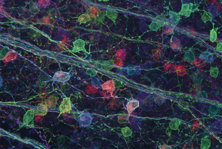
Now, thanks to Michael Fox and his team at the Virginia Tech Carilion Research Institute, we know that retina-to-brain connections don’t work quite the way neuroscientists previously thought. The researchers used a technique called “brainbow” (Figure 1), in which each individual neuron is tagged with a different fluorescent color, to trace what they assumed would be a single RGC – the source of several axon terminals forming synaptic connections. But when Aboozar Monavarfeshani, the graduate student who did the tagging, looked at the samples, he saw a variety of colors indicating terminals from more than just one or two RGCs. “The samples showed a true ‘brainbow’ – I could see, right in front of me, something very different than the concept I learned from my textbooks.” The researchers verified their results by electron microscopy and reached the same conclusion: that the axon terminals from numerous RGCs, rather than the few previously thought, make synaptic connections to the LGN (1). What does this mean for vision research? “These results are not what we expected, and they will force us to reevaluate our understanding of the architecture and flow of visual information through neural pathways,” said Fox. With such new and unanticipated information, scientists who seek to understand how the brain contributes to sight may have to reconsider theories that might once have been taken as read. But though these experiments raise more questions than they answer, it’s simply a sign that we have a long way to go to fully understand the complex visual machinery of the brain.

References

  1. S Hammer, et al., “Multiple retinal axons converge onto relay cells in the adult mouse thalamus”, Cell Rep, 12, 1575–1583 (2015). PMID: 26321636.

About the Author(s)

Michael Schubert

While obtaining degrees in biology from the University of Alberta and biochemistry from Penn State College of Medicine, I worked as a freelance science and medical writer. I was able to hone my skills in research, presentation and scientific writing by assembling grants and journal articles, speaking at international conferences, and consulting on topics ranging from medical education to comic book science. As much as I’ve enjoyed designing new bacteria and plausible superheroes, though, I’m more pleased than ever to be at Texere, using my writing and editing skills to create great content for a professional audience.

More Articles by Michael Schubert

Related Content

Newsletters

Receive the latest Ophthalmology news, personalities, education, and career development – weekly to your inbox.

Newsletter Signup Image

False

Advertisement

False

Advertisement

Explore More in Ophthalmology

Dive deeper into the world of Ophthalmology. Explore the latest articles, case studies, expert insights, and groundbreaking research.

False

Advertisement
The Ophthalmologist
Subscribe

About

  • About Us
  • Work at Conexiant Europe
  • Terms and Conditions
  • Privacy Policy
  • Advertise With Us
  • Contact Us

Copyright © 2025 Texere Publishing Limited (trading as Conexiant), with registered number 08113419 whose registered office is at Booths No. 1, Booths Park, Chelford Road, Knutsford, England, WA16 8GS.

Disclaimer

The Ophthalmologist website is intended solely for the eyes of healthcare professionals. Please confirm below: