Educational content provided by Alimera Sciences.
Diabetic macular edema (DME), the major cause of vision loss in patients with diabetic retinopathy, is characterized by vascular leakage, tissue edema, and the deposition of hard exudates in the central retina.1 Until the recent introduction of pharmacologic therapies, laser photocoagulation was the standard treatment. Despite the changes in treatment, the use of pharmacological treatments as single therapies mostly target vascular leakage and do not combat the underlying pathology.2 Thus, the management of DME remains complex, and often multiple treatment approaches are needed to resolve the persistence of fluid in the macula.
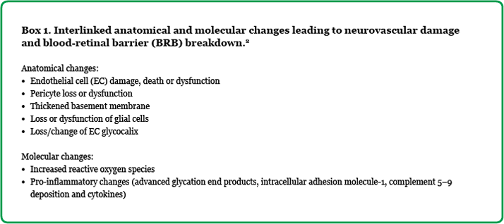
Pathophysiology of DME In diabetes mellitus (types 1 and 2) hyperglycemia causes overproduction of reactive oxygen species (ROS) in the mitochondria of cells within the neurovascular unit (endothelial cells, pericytes, astrocytes, and microglia), which leads to neurovascular damage and blood-retinal barrier (BRB) breakdown through a number of anatomical and molecular changes (Box 1).2,3 Blood-retinal barrier breakdown allows passage of both small and large molecules, and water through the BRB, causing a change in hydrostatic and oncotic pressure. This leads to an accumulation of fluid within the intraretinal layers of the macula and, ultimately, macular edema.4,5

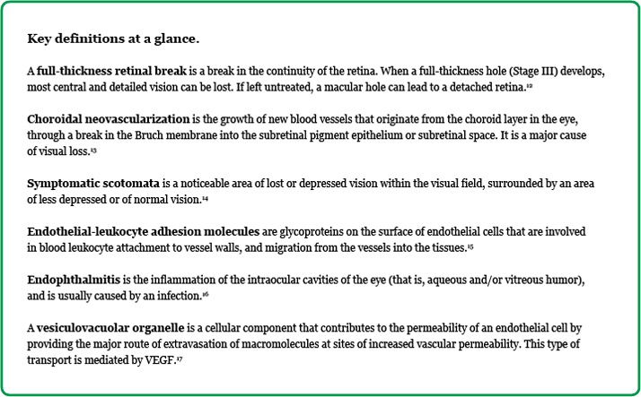
Current approaches to a disease with multifactorial etiology Laser photocoagulation The mechanism of action of laser photocoagulation-induced resolution of DME is poorly understood; however, one possible explanation is that oxygen-consuming photoreceptors are destroyed by the laser. Subsequently, oxygen can now diffuse through the laser scar tissue to the inner retina, relieving hypoxia of the inner retina.5 This form of therapy can lead to complications, such as full-thickness retinal break, choroidal neovascularization, subretinal fibrosis or symptomatic scotomata; all of which can lead to symptomatic visual loss.5 Despite this, laser photocoagulation is still the preferred therapy in some clinical settings for the treatment of DME because it is cost effective and requires less intensive management compared with some newer treatment modalities.4 Anti-VEGF therapy Expression of vascular endothelial growth factor (VEGF) is increased in DME due to factors such as hypoxia and hyperglycemia.4,6 VEGF induces phosphorylation of both tight junction proteins and an endothelial cell-cell adhesion molecule, leading to increased vascular permeability. VEGF also induces formation of vesiculovacuolar organelles (VVOs) within endothelial cells which can form transendothelial cell pores, allowing passage of large molecules and fluid through endothelial cells.4 These mechanisms directly increase vascular permeability and cause breakdown of the BRB. Anti-VEGFs—such as pegaptanib, ranibizumab, aflibercept, and bevacizumab—bind to portions of some or all isoforms of VEGF, inhibiting VEGF action, and are used for the treatment of age-related macular degeneration. However, several potential adverse effects have been linked to anti-VEGF therapy and the frequency of injections, including endophthalmitis, traumatic injury of the lens, and retinal detachment (risk rate <1.5% per patient per year).7 Corticosteroids In diabetes, there is increased expression of leukocyte adhesion molecules on the endothelial cell walls.6 Leukocytes release free radicals and enzymes that cause direct damage to endothelial cells, increasing BRB permeability. They also release cytokines (such as vascular endothelial growth factor [VEGF], tumor necrosis factor-α [TNF-α] and interleukin-6 [IL-6]), which act through various signaling pathways to increase vascular permeability.4 Inhibiting release of these cytokines with corticosteroids—such as dexamethasone, triamcinolone or fluocinolone—has been investigated within several clinical studies, and they have been shown to reduce macular edema. In particular, fluocinolone intravitreal implant 0.2 µg/day has been shown to be more effective in eyes with long-standing DME, where chronic inflammation was more important than the VEGF-mediated edema.2,8 However, a risk of elevated intraocular pressure has also been associated with corticosteroid DME therapy.2 Intraocular sustained-release corticosteroid devices are now commonly implanted or inserted into the eye, reducing patient discomfort and reducing the risk of the potential side effects of intravitreous injections, such as endophthalmitis and vitreous hemorrhage.9
Combination therapies that may be clinically effective In DME, significant changes in the cytokine profile have been reported, with elevated levels of VEGF, IL-6 and intercellular adhesion moleculE-1 (ICAM-1) among others. These changes result in chronic subclinical inflammation, which is important in the pathogenesis of the disease. It has been suggested that anti-VEGF drugs may only affect VEGF levels in intraocular fluids.2 It is therefore implied that targeting VEGF, or VEGF-independent pathways in isolation may not adequately block the increased permeability associated with diabetes and hyperglycemia.2 Corticosteroids suppress inflammation of retinal endothelial cells (RECs) by regulating inflammatory mediators, as well as by directly affecting ECs by regulating the phosphorylation, organization, and content of tight junction (TJ) proteins; inhibiting TJ disassembly and breakdown of the BRB.2 It then follows that steroids and anti-VEGFs may have complementary and additive effects on retinal vascular permeability, by both inhibiting the breakdown of the BRB barrier and suppressing inflammation.2 Dexamethasone (DEX) and anti-VEGF combination therapy may allow faster visual acuity (VA) improvements with a longer remission of DME.2 The DEX implant only lasts three to six months, therefore, any associated intraocular pressure (IOP) rise is short-lived.2 Furthermore, a recent trial of DEX intravitreal implant showed that relatively few implant injections would be required on average over three years (4.1, 4.4, and 3.3 for DEX implant 0.7 mg, DEX implant 0.35 mg and sham, respectively).10 Thus, the reduced frequency of intravitreal anti-VEGF injections would reduce the systemic adverse events associated with frequent anti-VEGF therapy. Fluocinolone (EU licensed dose, 0.2 µg/day)—a longer acting corticosteroid—may also play a role in combination with anti-VEGF therapy in phakic and pseudophakic eyes, as it is more effective in eyes with a longer duration of DME, where inflammation is the major cause of edema, despite the adverse event profile of corticosteroids, which includes cataracts and increased IOP.2,11

Laser treatment can lead to reduced oxygen consumption and induce a complex action on the retinal pigment epithelium.6 It has been shown that laser treatment combined with corticosteroids or anti-VEGF therapy can lead to better VA outcomes and reduced central macular thickness.6

Conclusion The management of DME remains complex, and often multiple treatment approaches are required to resolve the persistence of fluid within the macular region. Combination treatment of anti-VEGF therapies with corticosteroids may play an important role in the management of DME, and provide more optimal pharmacotherapy, supplemented with macular laser photocoagulation as necessary in individual cases. The ideal treatment for DME should be well tolerated by patients, cost effective, result in improved morphology of the macula (a completely dry macula), and improve vision for a significant duration with reduced adverse events.2

Conflict of interest: Winfried Amoaku has provided consultancy services to Alcon, Alimera Sciences, Allergan, Bayer, Novartis, Santen and Thrombogenics. He has received travel grants from Alimera Sciences, Allergan, Bayer and Novartis, and honoraria for lectures from Allergan and Novartis. He has participated in clinical trials for which his institution has received funding from Allergan, Novartis and Pfizer. His institution has further received research grants from Allergan and Novartis for non-clinical studies, and CentreVue (Italy) for clinical studies.

Look out for DME content developed by Alimera Sciences on this website throughout 2015. We hope it supports your knowledge of DME, and if you would like to contribute material for publication, please send your materials to dmecontenthub@hayward.co.uk, we’d be very pleased to consider your contributions.

REFERENCES
- I Klaassen et al., “Molecular basis of the inner blood-retinal barrier and its breakdown in diabetic macular edema and other pathological conditions”, Prog Retin Eye Res, 34, 19–48 (2013). PMID: 23416119.
- W Amoaku et al., “A review of therapies for diabetic macular oedema and rationale for combination therapy”, Eye (advance online publications), 1–16 (2015). PMID: 26113500.
- F Giacco, M Brownlee, “Oxidative stress and diabetic complications”, Circ Res, 107, 1058–70 (2010). PMID: 21030723 (circres.ahajournals.org/content/107/9/1058.long).
- A Wenick, N Bressler, “Diabetic macular edema: current and emerging therapies”, Middle East Afr J Ophthalmol, 19(1), 4–12 (2012). PMCID: PMC3277023
- N Bhagat et al., “Diabetic macular edema: pathogenesis and treatment”, Sur Ophthalmol 54 (1) January_February 2009 PMID: 19171208.
- S Rashaed, J Arevalo, “Combined therapy for diabetic macular edema”, Middle East Afr J Ophthalmol, 20(4), 315–320 (2013). PMCID: PMC3841949.
- R Simó, C Hernández, “Intravitreous anti-VEGF for diabetic retinopathy: hopes and fears for a new therapeutic strategy”, Diabetologia, 51, 574–580 (2008). PMID:18404258.
- PA Campochiaro et al., “Sustained delivery fluocinolone acetonide vitreous inserts provide benefit for at least 3 years in patients with diabetic macular edema”, Ophthalmology, 119(10), 2125–2132 (2012). PMID: 22727177.
- M Cabrera et al., “Sustained-Release Corticosteroid Options”, J Ophthalmol, 2014, 1–5 (2014). PMID:25140246 (www.hindawi.com/journals/joph/2014/164692/).
- DS Boyer et al., “Three-year, randomized, sham-controlled trial of dexamethasone intravitreal implant in patients with diabetic macular edema”, Ophthalmology, 121(10), 1904–1914 (2014). PMID: 24907062.
- Y Yang et al., “Long-term outcomes of phakic patients with diabetic macular oedema treated with intravitreal fluocinolone acetonide (FAc) implants”, Eye, 29, 1173–1180 (2015). PMID: 26353919 (http://www.nature.com/eye/journal/v29/n9/full/eye201598a.html).
- National Eye Institute (NEI), “Full-thickness retinal break”, (2015). Available at: nei.nih.gov/health/macularhole/macularhole. Accessed September 2015.
- Medscape, “Choroidal neovascularization”, (2015). Available at: emedicine.medscape.com/article/1190818-overview. Accessed September 2015.
- The Medical Dictionary, The free dictionary by Farlex, “Scotomata”, (2015). Available at: medical-dictionary.thefreedictionary.com/scotoma. Accessed September 2015.
- The Medical Dictionary, The free dictionary by Farlex, “Endothelial-leukocyte adhesion molecule”, (2015). Available at: medical-dictionary.thefreedictionary.com/endothelial-leukocyte+adhesion+molecule. Accessed September 2015.
- Medscape, “Endophthalmitis”, (2015). Available at: emedicine.medscape.com/article/799431-overview. Accessed September 2015.
- AM Dvorak, D Feng, “The vesicular–vacuolar organelle (VVO): a new endothelial cell permeability organelle”, J Histochem Cytochem, 49(4), 419–432 (2011). PMID: 11259444 (http://jhc.sagepub.com/content/49/4/419.long)
Founded in 2003, Alimera Sciences researches and develops innovative vision-improving treatments for chronic retinal diseases, such as diabetic macular edema (DME), dry age-related macular degeneration (AMD), and retinal vein occlusion. In 2015, Alimera Sciences partnered with The Ophthalmologist to facilitate the publication of independently created educational content surrounding DME, a serious retinal complication associated with diabetes, which is increasing in incidence with the increasing prevalence of diabetes worldwide. Published content will include articles ranging from basic science and disease processes to overviews of clinical data, different surgical procedures, comparisons of treatment options, and practical advice for managing diabetic patients. With a commitment to honesty, integrity, responsibility, candor, and trust, Alimera Sciences intend to provide educationally focused content to healthcare professionals across a wide range of topics in DME in order to both increase disease awareness and understanding, and to help improve patient outcomes. UK-ILV-MMM-0355 Date of preparation: August 2015 enquiries@alimerasciences.com

