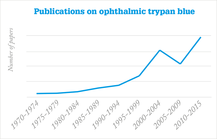
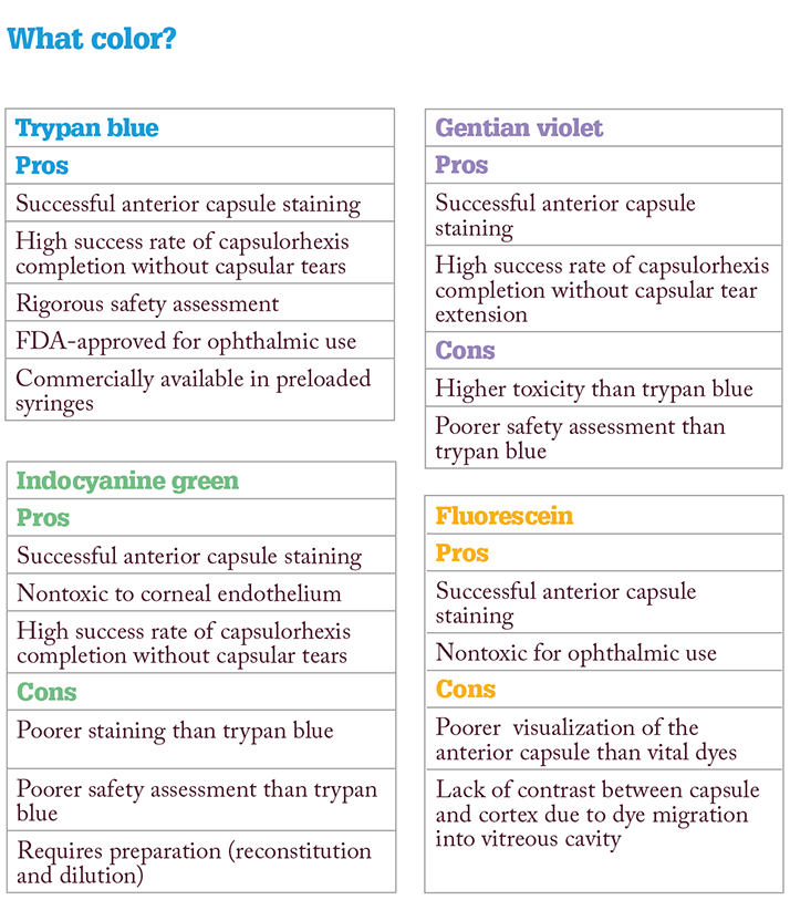
Vital dyes are an increasingly useful component of ophthalmic operations. Used to stain the anterior capsule for tissue identification, these stains are especially important when the view through the cornea is limited – for instance, in cases of corneal scarring, dense cataract or compromised red fundus reflex. Trypan blue is the most popular of the vital dyes – but how did it reach its current popularity, and what should ophthalmologists know about using it?
Vital do’s and vital don’ts
- For cataract surgery, stain the lens capsule whenever view through the cornea is compromised or there is dens cataract, poor red reflex or small pupil
- When staining the anterior capsule, wait 20–30 seconds after application
- For donor tissue preparation during deep anterior lamellar keratoplasty, stain the donor Descemet’s membrane to ensure complete removal prior to suturing the host cornea
- When staining for Descemet’s membrane endothelial keratoplasty, wait 2–3 minutes after application for complete graft staining
- The improved visualization offered by vital dyes may be useful for surgeon training
- Do not use vital dyes with silicone or acrylic intraocular lenses
A history of dyes
1904 - First synthesis of trypan blue by Paul Ehrlich (1)1972 - First perioperative trypan blue staining of corneal endothelium (2)
1996 - First intraoperative indocyanine green staining of anterior capsule (3)
1997 - First intraoperative fluorescein staining of anterior capsule (4)
1997 - First intraoperative trypan blue staining of anterior capsule (5)
1999 - CE Marking of ready-to-use trypan blue for anterior capsule staining (6)
2004 - FDA approval of ready-to-use trypan blue for anterior capsule staining
References
- ME Farah, et al., “Current concepts of trypan blue in chromovitrectomy”, Vital Dyes in Vitreoretinal Surgery, 91–100. Karger: 2008. MS Norn, “Per operative trypan blue vital staining of corneal endothelium. Eight years’ follow up”, Acta Ophthalmol (Copenh), 58, 550–555 (1980). PMID: 6163309. M Horiguchi, et al., “Staining of the lens capsule for circular continuous capsulorrhexis in eyes with white cataract”, Arch Ophthalmol, 116, 535–537 (1998). PMID: 9565058. WL Fritz, “Fluorescein blue, light-assisted capsulorhexis for mature or hypermature cataract”, J Cataract Refract Surg, 24, 19–20 (1998). PMID: 9494894. GR Melles, et al., “Trypan blue capsule staining to visualize the capsulorhexis in cataract surgery”, J Cataract Refract Surg, 25, 7–9 (1999). PMID: 9888070. DF Chang, “Trypan blue versus indocyanine green”, (2005). Available at: http://bit.ly/1kf0Kj4. Accessed October 29, 2015. R Daly, “Surgeons beware: the wrong dye leads to edema and discoloration”, (2007). Available at: http://bit.ly/1WkwkNI. Accessed October 29, 2015.
