Sometimes, slit lamp examination of blebs following trabeculectomy isn’t sufficient to evaluate bleb function. You can visualize the bleb and compare what you see with a number of pre-defined bleb type classifications, such as such as cystic, encysted, flat, and diffuse… but although these terms may be useful for describing common appearances, they certainly aren’t perfect. Not only do they lack the depth of description required for following bleb morphologic changes over time, but there is also a vast range of possible variations within each of these descriptions (1).
OCT imaging, in vivo confocal microscopy and ultrasound biomicroscopy (UBM) have all been employed to examine the internal structures of the filtering bleb, but there are drawbacks associated with these approaches too – for example, UBM is a contact technique (and risks bleb-related complications), and OCT imaging can’t replace the slit lamp for the assessment of bleb vascularity and Seidel status. Thermography has previously shown promise as an alternative method of bleb assessment (2). Aqueous humor is cooler than surrounding tissue, so as it flows out of the scleral flap, it cools the subconjunctival space it fills. Poorly functioning blebs will produce less of a cooling effect. Based on that premise, a team from the Department of Ophthalmology at Charité-University Medicine Berlin decided to evaluate filtering bleb function in 35 eyes of 35 patients with primary open-angle glaucoma who underwent trabeculectomy using a new non-contact ocular surface thermography device (1).
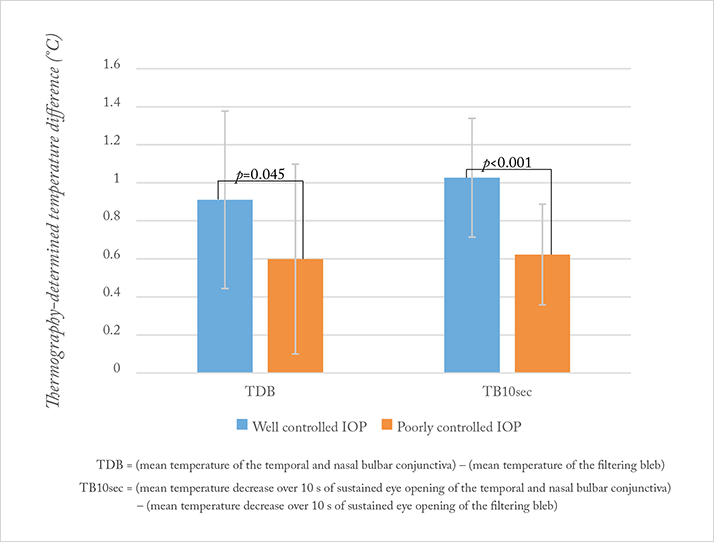
Patients were divided into two groups: well-controlled IOP (<18 mmHg, no glaucoma medications required) and poorly-controlled IOP (≥18 mmHg, ≥1 glaucoma medication required). All eyes were assessed with the thermographer in order to derive two key parameters: the mean temperature decrease in the filtering bleb at a single timepoint (TDB), and the same over a period of 10 seconds of sustained eye opening (TB10sec; Figure 1). Both parameters were found to be significantly different in patients with well-controlled IOP versus poorly controlled IOP (Figure 1). If these findings hold true in larger studies, thermography could offer a noninvasive and relatively inexpensive tool to assess bleb function.
References
- MKJ Klamann, et al., “Thermography: a new option to monitor filtering bleb function?”, J Glaucoma, 24, 272–277 (2015). PMID: 23708421. S Kawasaki, et al., “Evaluation of filtering bleb function by thermography”, Br J Opthalmol, 93, 1331–1336 (2009). PMID: 19520695.
