Conexiant
Login
  • Corneal Physician
  • Glaucoma Physician
  • New Retinal Physician
  • Ophthalmology Management
  • Ophthalmic Professional
  • Presbyopia Physician
  • Retinal Physician
The Ophthalmologist
  • Explore

    Explore

    • Latest
    • Insights
    • Case Studies
    • Opinion & Personal Narratives
    • Research & Innovations
    • Product Profiles

    Featured Topics

    • Anterior Segment
    • Glaucoma
    • Retina

    Issues

    • Latest Issue
    • Archive
  • Subspecialties
    • Cataract
    • Cornea
    • Glaucoma
    • Neuro-ophthalmology
    • Oculoplastics
    • Pediatric
    • Retina
  • Business

    Business & Profession

    • Professional Development
    • Business and Entrepreneurship
    • Practice Management
    • Health Economics & Policy
  • Training & Education

    Career Development

    • Professional Development
    • Career Pathways

    Events

    • Webinars
    • Live Events
  • Events
    • Live Events
    • Webinars
  • Community

    People & Profiles

    • Power List
    • Voices in the Community
    • Authors & Contributors
  • Multimedia
    • Video
    • Podcasts
Subscribe
Subscribe

False

Advertisement
The Ophthalmologist / Issues / 2015 / Mar / Optic Ultrasound Predicts Stroke Outcomes
Neuro-ophthalmology

Optic Ultrasound Predicts Stroke Outcomes

After a stroke, the brain swells... and for every millimeter that the optic nerve sheath diameter swells, the risk of death increases by 4–6 times.

By Mark Hillen 3/17/2015 1 min read

Share

0315-205-main

Imaging the retina to understand systemic disease is not a new concept – you’re able to noninvasively image not only the retinal vasculature, but with optic nerve and the retinal nerve fiber layer, neural tissue too. This is beginning to be exploited to map the progression of both vascular and neurological diseases (1,2). It’s also starting to be useful in predicting outcomes after patients have experienced a stroke (3).

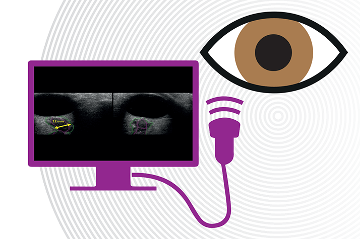
Severe strokes tend to result in sharp increases in intracranial pressure (ICP) – and makes for a poor prognosis for the patient. ICP can be measured by lumbar puncture, but this is invasive, and can easily be confounded by other aspects of the patient’s medical management, such as acute sedative use. It turns out that measuring the optic nerve sheath diameter (ONSD) with optic ultrasonography (OUS) is a promising method of assessing ICP.

How was this established? Researchers at the Gainesville campus at the University of Florida performed OUS on patients (n=86) who presented with stroke on both the day of admission and one day later, measuring ONSD – transversely and laterally – in both eyes (3). Regression analysis was then used to assess the relationship between ONSD and patient outcomes.

0315-205-fig1Figure 1. a. Odds ratio of mortality 6 months after stroke; b. Mean in-hospital ONSD was smaller in patients who had survived at 6 months post-stroke, compared with those who died within that period. ONSD, optic nerve sheath diameter; ICH, intracerebral hemorrhage; IS, ischemic stroke.

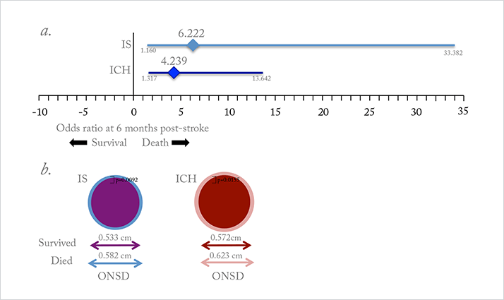
The researchers found that there was a significant difference in mean ONSD between patients who died within the first six months and those who survived: for every extra millimeter, the risk of death was four times higher in patients with ischemic stroke, and six times higher in patients with intracerebral hemorrhage (Figure 1a). Larger ONSD was also associated with a greater probability of death after six months (Figure 1b), poorer functional outcomes, and more severe disability. The authors are hopeful that OUS could offer an attractive screening alternative for ICP, as it is a routine bedside test that is performed using gel and a device placed on closed eyelids, potentially allowing doctors to reliably and noninvasively detect elevated ICP and initiate ICP-lowering treatment earlier.

References

  1. M Zacharria, “In the Eye of the Storm”, The Ophthalmologist, 1, 16–23 (2013). http://bit.ly/top0113. WW Lee et al., “Retinal nerve fiber layer structure abnormalities in schizophrenia and its relationship to disease state: evidence from optical coherence tomography”, Invest Ophthalmol Vis Sci, 54, 7785–7792 (2013). PMID: 24135757. VS Hedna et al., “Use of optic nerve sheath diameter in emergency department to predict stroke outcome”, Stroke, 46, Abstract: AWMP83 (2015).

About the Author(s)

Mark Hillen

I spent seven years as a medical writer, writing primary and review manuscripts, congress presentations and marketing materials for numerous – and mostly German – pharmaceutical companies. Prior to my adventures in medical communications, I was a Wellcome Trust PhD student at the University of Edinburgh.

More Articles by Mark Hillen

Related Content

Newsletters

Receive the latest Ophthalmology news, personalities, education, and career development – weekly to your inbox.

Newsletter Signup Image

False

Advertisement

False

Advertisement

Explore More in Ophthalmology

Dive deeper into the world of Ophthalmology. Explore the latest articles, case studies, expert insights, and groundbreaking research.

False

Advertisement
The Ophthalmologist
Subscribe

About

  • About Us
  • Work at Conexiant Europe
  • Terms and Conditions
  • Privacy Policy
  • Advertise With Us
  • Contact Us

Copyright © 2025 Texere Publishing Limited (trading as Conexiant), with registered number 08113419 whose registered office is at Booths No. 1, Booths Park, Chelford Road, Knutsford, England, WA16 8GS.

Disclaimer

The Ophthalmologist website is intended solely for the eyes of healthcare professionals. Please confirm below: