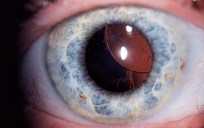
Photographer: Terry Cooper, Volk Optical.
The Vision 2020 Links team – a group of ophthalmologists, orthoptists, and nurses from the Royal Free Hospital in London led by consultant ophthalmologist Clare Davey – went on a 10-day mission to Uganda in April. Photographs documenting that visit are inside this section.
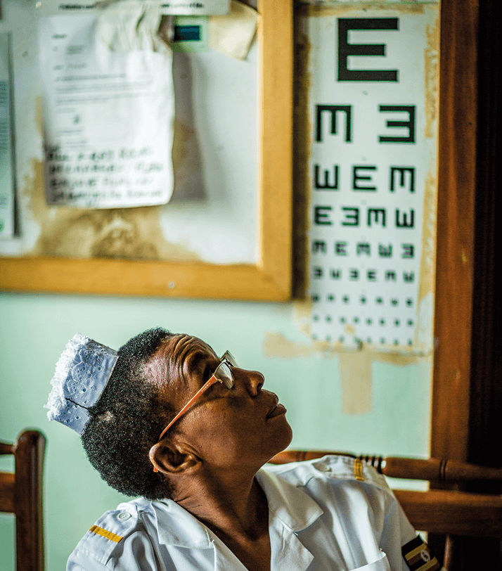
Photographer: Terry Cooper, Volk Optical.
Checking a diabetic patient for eye disease at Mulago Hospital, Kampala, Uganda.
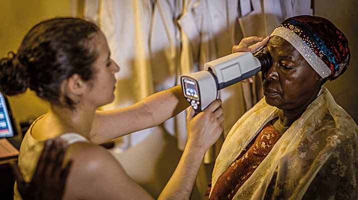
Laser treatment of a patient with diabetic eye disease.
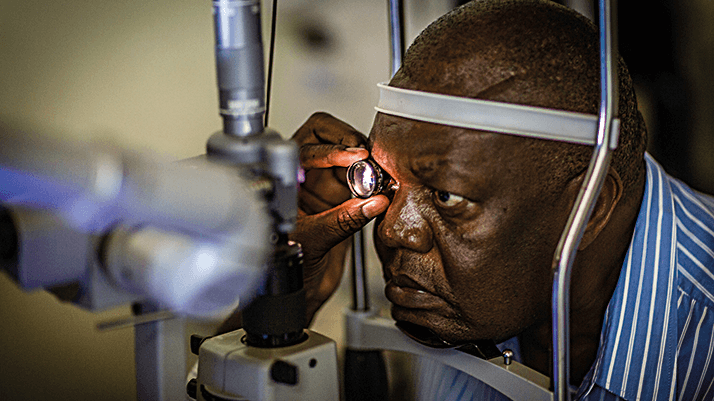
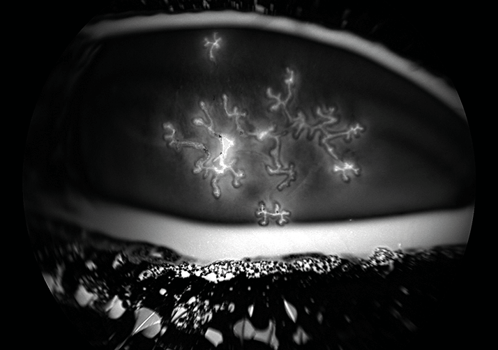
Image credit: Zachary Dupureur & David A Johnson
Autofluorescence of bilateral herpetic dendritic keratitis, dyed using fluorescein strips.
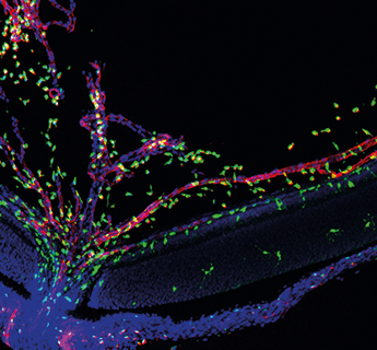
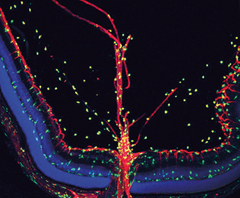
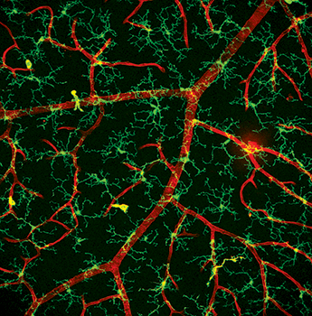
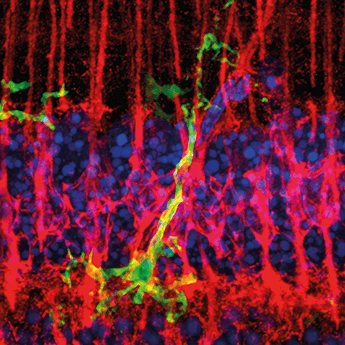
Wai Wong, Chief of the Unit on Neuron-Glia Interactions in Retinal Disease, National Eye Institute, National Institutes of Health.
a. Microglia (green) migrating into the developing retina at postnatal day 3 via the developing retinal vasculature and hyaloid vessels (red).
b. Microglia (green) migrating into the developing retina at postnatal day 7.
c. Microglia cells (green) in an en-face view of the inner plexiform layer in close proximity to retinal capillaries (red).
d. Microglial cell (green) fasciculating closely with radial Müller cell processes (red) in an endotoxin model of retinal inflammation.
Photographer: Bohdan Kousal, Department of Ophthalmology, Charles University, Prague, Czech Republic
A superiorly subluxated intraocular lens.
