Conexiant
Login
  • Corneal Physician
  • Glaucoma Physician
  • New Retinal Physician
  • Ophthalmology Management
  • Ophthalmic Professional
  • Presbyopia Physician
  • Retinal Physician
The Ophthalmologist
  • Explore

    Explore

    • Latest
    • Insights
    • Case Studies
    • Opinion & Personal Narratives
    • Research & Innovations
    • Product Profiles

    Featured Topics

    • Anterior Segment
    • Glaucoma
    • Retina

    Issues

    • Latest Issue
    • Archive
  • Subspecialties
    • Cataract
    • Cornea
    • Glaucoma
    • Neuro-ophthalmology
    • Oculoplastics
    • Pediatric
    • Retina
  • Business

    Business & Profession

    • Professional Development
    • Business and Entrepreneurship
    • Practice Management
    • Health Economics & Policy
  • Training & Education

    Career Development

    • Professional Development
    • Career Pathways

    Events

    • Webinars
    • Live Events
  • Events
    • Live Events
    • Webinars
  • Community

    People & Profiles

    • Power List
    • Voices in the Community
    • Authors & Contributors
  • Multimedia
    • Video
    • Podcasts
Subscribe
Subscribe

False

Advertisement
The Ophthalmologist / Issues / 2015 / Jun / Images of Ophthalmology

Images of Ophthalmology

Popular culture has always viewed eyes as things of beauty. But there’s more to the eye than the color of your irides. Advances in imaging technology have enabled us to see far more than could ever have been imagined. And on closer inspection, they’re still beautiful.

By Mark Hillen, Roisin McGuigan, Michael Schubert 6/8/2015 1 min read

Share

Iris Vascular Malformation. John McCormick, Tennent Institute of Ophthalmology, Glasgow, UK.

Supported by

By John McCormick, photographed with the Haag-Streit BX 900 Slit Lamp.

Ophthalmic Diagnostic & Clinical Imaging, Tennent Institute of Ophthalmology Gartnavel General Hospital, Glasgow, Scotland.

Matt Poe is a certified ophthalmic assistant working towards certification in retinal angiography. He runs the ophthalmic imaging site www.ophthalmicphotography.info.

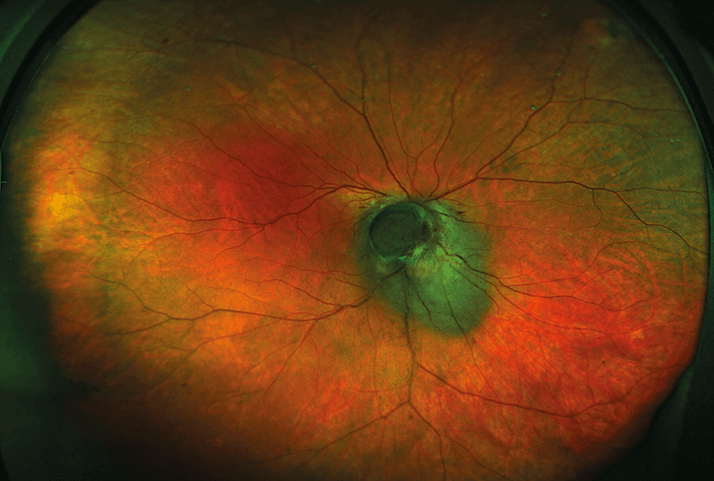
This patient was first diagnosed with a choroidal melanocytoma. The patient did not return to the clinic for three years until one month when he lost vision on and off several times a day. The flat melanocytoma had progressed to a large, mushroom-shaped tumor. The tumor’s location meant that radiotherapy was contraindicated. Enucleation was performed, and the tumor was confirmed to be uveal melanoma.

Amra Nadarevic Vodencarevic, Resident Ophthalmologist, University Clinic Center Tuzla, Bosnia and Herzegovina.

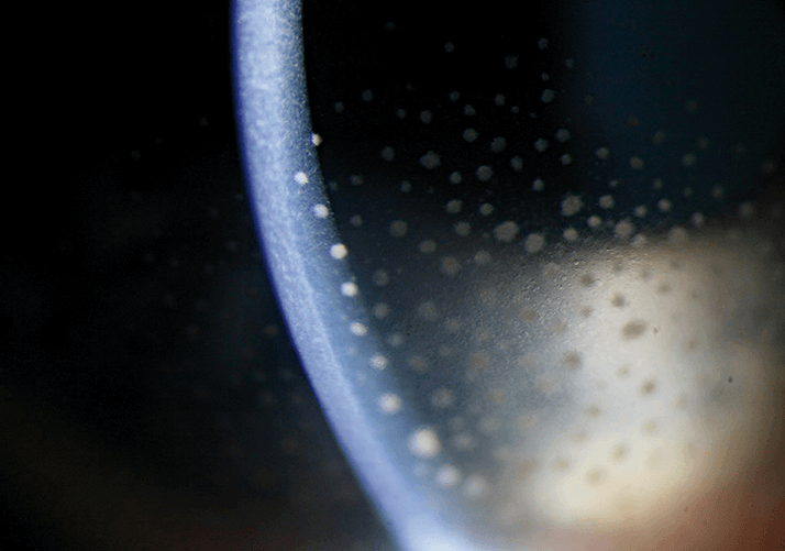
Bubble of silicon in the anterior chamber.

Angela Chappell is an ophthalmic photographer at Flinders Medical Centre, Adelaide, South Australia. Images copyright: Flinders Centre of Ophthalmology.

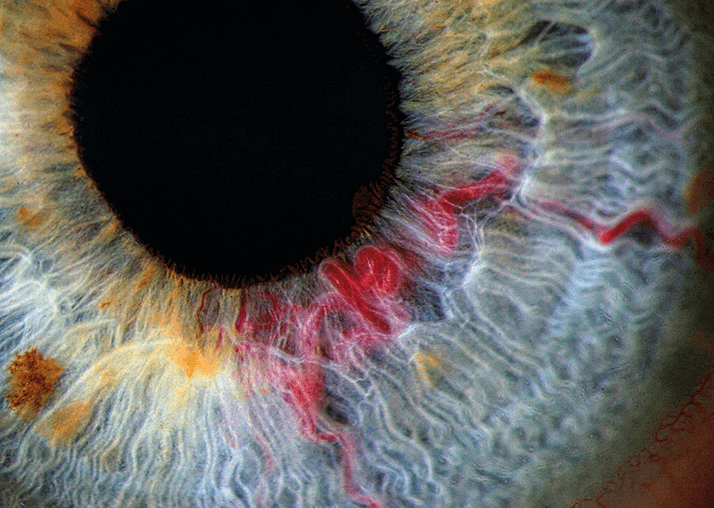
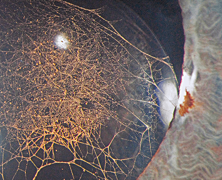
Fibrin web formation with pigment deposition on a decentered posterior chamber lens implant.

Helena Prior Filipe is a consultant ophthalmologist at the Instituto Dr Gama Pinto, Lisbon, Portugal

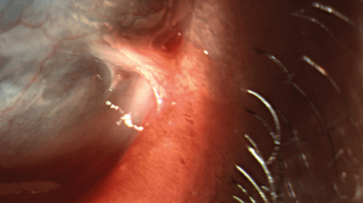
Simblepharon in a Lyell syndrome case: lower eyelid and inferior bulbar conjunctival adhesion.

Tarun Arora, senior resident, the Rajendra Prasad Centre for Ophthalmic Sciences, All India Institute of Medical Sciences, New Delhi, India.

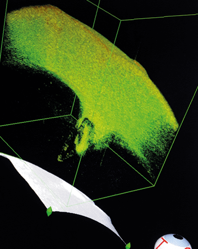
Anterior segment OCT of glass penetrating the anterior chamber.

Carrie A. Cooke is an ophthalmic photographer with the University of Texas Health Science Center San Antonio, Medical Arts and Research Center, Department of Ophthalmology.

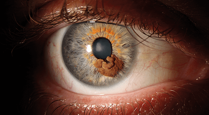
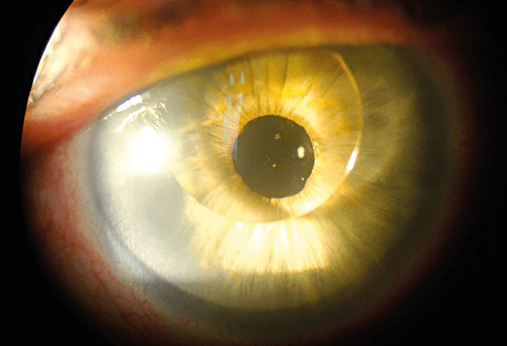
This patient presented to our clinic after a routine exam with a shopping center optometrist. The patient explained that the growth had been present since he was an infant and had changed very little throughout his life. The diagnosis was a non-malignant congenital anomaly of the iris and will be monitored every 6 months.

Images of the Posterior Segment
Incredible Images

About the Author(s)

Mark Hillen

I spent seven years as a medical writer, writing primary and review manuscripts, congress presentations and marketing materials for numerous – and mostly German – pharmaceutical companies. Prior to my adventures in medical communications, I was a Wellcome Trust PhD student at the University of Edinburgh.

More Articles by Mark Hillen

Roisin McGuigan

I have an extensive academic background in the life sciences, having studied forensic biology and human medical genetics in my time at Strathclyde and Glasgow Universities. My research, data presentation and bioinformatics skills plus my ‘wet lab’ experience have been a superb grounding for my role as a deputy editor at Texere Publishing. The job allows me to utilize my hard-learned academic skills and experience in my current position within an exciting and contemporary publishing company.

More Articles by Roisin McGuigan

Michael Schubert

While obtaining degrees in biology from the University of Alberta and biochemistry from Penn State College of Medicine, I worked as a freelance science and medical writer. I was able to hone my skills in research, presentation and scientific writing by assembling grants and journal articles, speaking at international conferences, and consulting on topics ranging from medical education to comic book science. As much as I’ve enjoyed designing new bacteria and plausible superheroes, though, I’m more pleased than ever to be at Texere, using my writing and editing skills to create great content for a professional audience.

More Articles by Michael Schubert

Related Content

Newsletters

Receive the latest Ophthalmology news, personalities, education, and career development – weekly to your inbox.

Newsletter Signup Image

False

Advertisement

False

Advertisement

Explore More in Ophthalmology

Dive deeper into the world of Ophthalmology. Explore the latest articles, case studies, expert insights, and groundbreaking research.

False

Advertisement
The Ophthalmologist
Subscribe

About

  • About Us
  • Work at Conexiant Europe
  • Terms and Conditions
  • Privacy Policy
  • Advertise With Us
  • Contact Us

Copyright © 2025 Texere Publishing Limited (trading as Conexiant), with registered number 08113419 whose registered office is at Booths No. 1, Booths Park, Chelford Road, Knutsford, England, WA16 8GS.

Disclaimer

The Ophthalmologist website is intended solely for the eyes of healthcare professionals. Please confirm below: