Florian Kretz Eye Clinic Ahaus-Raesfeld Rheine, Ahaus, Germany
Keith Barton Moorfields Eye Hospital, London, UK
Kuldev Singh Stamford School of Medicine, Palo Alto, California, USA
Michael Koss Goethe University, Frankfurt am Main, Germany
Michael Mrochen IROC Science, Zürich, Switzerland
Bill Trattler Center for Excellence in Eye Care, Miami, Florida, USA
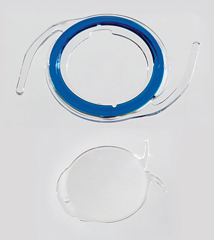
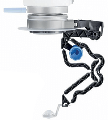
A two-piece IOL designed to adapt to, and accommodate changes in, refractive needs
Produced by: ClarVista Medical, Inc.Detail: The HARMONI modular IOL system is designed to improve upon existing IOL designs, by having the flexibility to exchange the optic component at any time – without manipulation of the capsular bag or the base component. HARMONI’s base component enhances structural support of the capsular bag, in order to help deliver more predictable post-procedural outcomes. The base component is designed to securely receive the optic component using traditional surgical tools and techniques, and the optic component can be of monofocal, toric, advanced multifocal or extended depth-of-focus designs – and can be exchanged post-operatively without manipulation of the base or the delicate capsular bag. The two-piece design means that the base plate has the potential to maintain a consistent position within the capsular bag – and that the addition of the optic portion of the IOL to the base plate should attain a more predictable position within the eye. The effective lens position can therefore be better predicted, allowing for improved outcomes. If the desired power is missed, the fact that the optic portion of the IOL can be easily exchanged allows for a safer and more predictable enhancement than current IOL designs permit. In the case of toric misalignment, realignment should also be safer and easier than current IOL designs. Impact: The HARMONI Modular IOL system is designed to produce a more predictable lens position, and allow for refractive care to be provided over the patient’s life, even if changes in lifestyle or pathology create the need for a different refractive solution, as it will allow patients to upgrade their IOL as better technologies arise. The HARMONI allows for easy adjustment of IOL power, addition (or subtraction) of toricity, IOL rotation by exchange, or manipulation of the modular component of the optic in these two-piece component IOLs. It is also potentially very important for pediatric cataract patients, who experience changes in eye power as the eye grows. The judges said:
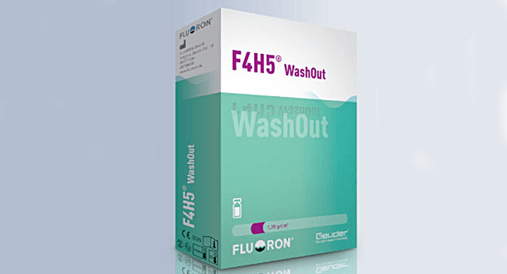
Amphiphilic surfactant for complications in silicone oil removal
Produced by: GeuderDetail: F4H5 WashOut (perfluorobutylpentane – C4F9-C5H11) is a semifluorinated alkane that is able to dissolve silicone oil. F4H5 WashOut is similar to another solvent for silicon oil, F6H8 (perfluorohexyloctane—C6F13- C8H17) but is more amphiphilic (i.e. both hydrophilic and lipophilic) and therefore a superior solvent for silicone oil. Crucially, F4H5 forms no potentially reactive structures, and should therefore ensure biocompatibility. F4H5 WashOut is able dissolve silicon oil in balanced salt solution at any mixing ratio – a situation where conventional surfactants have limited success. Impact: Tamponades like silicone oil and perfluorocarbon liquids (PFCL) are today’s gold standard in modern vitreoretinal surgery. But although they are necessary, and can positively impact clinical results, there are also potential side effects, such as emulsified oil, unwanted mixtures of silicone oil and PFCL, and residual oil that remains in the eye. This situation can entail unwanted clinical manifestations, including glaucoma, inflammation and formation of fibrosis, and proliferative vitreoretinopathy, so it’s important to eradicate silicon oil completely to avoid risking these complications. Increased use of PFCL and silicone oil, as well as increasingly smaller incision sizes (which can make it more complicated to completely remove the vitreous, as well as inducing tamponades) can increase the incidence of adverse effects – making a biocompatible, effective detergent a necessity. The judges said:
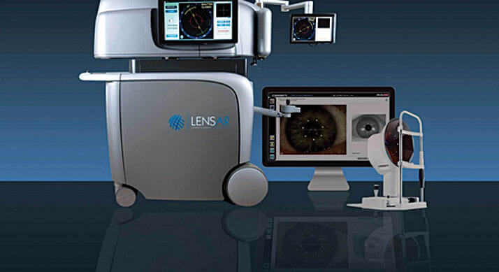
The first femto cataract platform to automate several key steps of the procedure
Produced by: LenSAR, Inc.Detail: The LENSAR laser system with Streamline is the first femtosecond cataract laser that can automate several key steps of surgical planning and delivery. It can integrate wirelessly with certain preoperative corneal topographers (like iOptics’ Cassini), and can perform iris registration, automatic cataract density imaging, and automatic customized fragmentation patterns. The system can perform anterior laser capsulotomies, lens fragmentation, and corneal and arcuate incisions, and its level of automation allows for integration into existing workflows without increasing procedure times. Impact: The integration and automation the system offers has the potential to reduce errors that can occasionally arise from manual data entry and the issues that can arise from physically marking the eye preoperatively. This should increase accuracy and efficiency (and potentially visual outcomes too). This is the first system to fully automate and customize these important planning and execution steps of refractive surgery The judges said:
A smartphone app to connect healthcare workers with ophthalmic guidelines and advice
Produced by: William Mapham, Ophthalmology registrar, University of Stellebosch, Tygerberg Hospital, South AfricaDetail: The Vula smartphone application provides healthcare workers with basic ophthalmic diagnostic guidelines, and connects them with an ophthalmologist who can provide advice, respond to queries, and accept referrals in real time. Vula has the potential to bring specialist eye care to anyone, anywhere. Aimed at rural communities where access to specialist ophthalmic care is extremely difficult to obtain, the Vula app empowers community healthcare workers with decision making assistance, and access to specialist knowledge via their mobile phones. By equipping healthcare workers with the ability to conduct a basic ophthalmic examination, complete a standardized referral form, and access a local specialist for consultation and referral in real time, it ensures that eye conditions are appropriately managed and referred. The app can be used anywhere in the world, connecting healthcare workers with their local ophthalmologist. Impact: Vula extends the reach of specialist eye care beyond the physical boundaries of the eye clinic. As a mobile platform, it can overcome the often poor traditional communication and transport infrastructure, taking advantage of the high penetration of mobile phones in even the poorest of communities. The judges said:

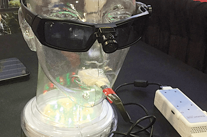
A nonsurgical intervention to let the visually impaired “see with their tongue”
Produced by: Wicab Inc.Detail: The BrainPort V100 is an oral electronic vision aid that provides electro-tactile stimulation to aid profoundly blind patients in orientation, mobility, and object recognition as an adjunctive device to other assistive methods, such as the white cane, or a guide dog. The technology translates digital information from a wearable video camera into gentle electrical stimulation patterns on the surface of the tongue. Users feel moving bubble-like patterns on their tongue which they learn to interpret as the shape, size, location and motion of objects in their environment. Some users have described it as being able to “see with your tongue.” Impact: The BrainPort V100 is a nonsurgical intervention that can be used by individuals with no vision, irrespective of whether they are congenitally blind, or have acquired blindness. The technology could allow blind people who cannot currently be treated to live more independently – and as BrainPort does not affect the eyes, this could be beneficial if future research offers better surgical or other therapeutic alternatives. One judge said:
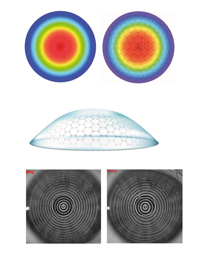
SmartPulse technology helps perfects corneal smoothness during corneal ablation, to optimize short-term clinical outcomes
Produced by: SCHWIND eye-tech-solutionsDetail: SmartPulse uses a sophisticate three-dimensional model – based on the structure of fullerenes – that realistically describes the curvature of the cornea, and which makes it possible to position the laser pulses more closely than has been achieved before. The latest measurement and analysis methods help make optimum use of the spot geometry. Using SmartPulse results in a very smooth stromal bed after the ablation process, with fast epithelial closure in surface treatments. This can enhance short-term outcomes by reducing residual roughness, and improves the smoothness of the residual bed without compromising the stability of long-term outcomes. SmartPulse technology improves patients’ visual acuity and quality in the early postoperative phase of all treatment methods, irrespective of whether flap technique, stromal, or surface ablation is used. The effect of a very smooth corneal surface is most evident with surface treatments, where neither a LASIK flap nor epithelium helps smooth the stromal surface before regeneration. Impact: A recent multi-center evaluation with 1,000 eyes illustrated that SmartPulse provides excellent results, particularly in the early postoperative stage. All patients underwent TransPRK and the eight international surgeons involved in the study reported shorter recovery time of visual acuity, higher levels of postoperative visual quality, and shorter re-epithelialization in the patients they treated. One judge said:
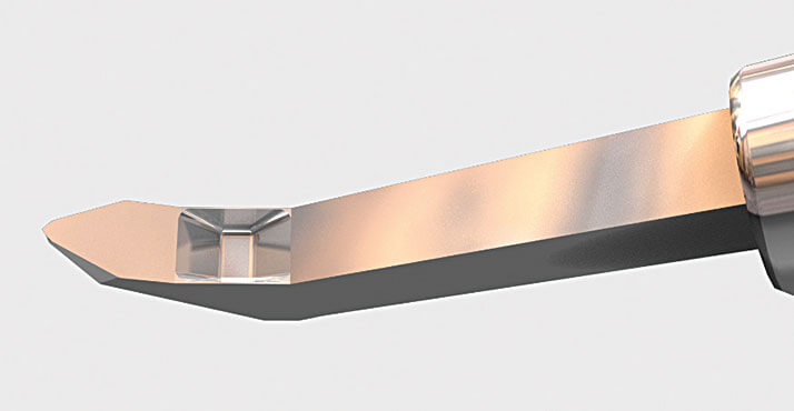
Micro-machined parallel blades designed to cleanly extract trabecular meshwork
Produced by: New World MedicalDetail: The Kahook Dual Blade (KDB) is designed to precisely remove trabecular meshwork in complete strips, to allow for aqueous drainage through the natural outflow system of the eye. The device will help treat glaucoma, and simplify the collection of trabecular tissue for study. Using precision micromachining and laser-cutting technology, the KDB is made from stainless steel, and engineered to excise trabecular meshwork (TM) tissue through a clear corneal incision as small as 1.2 mm. It features a tip to pierce the TM, a ramp that stretches as the KDB is advanced, and parallel blades that excise the stretched tissue. The heel is also precision engineered with rounded edges, in order to avoid damaging the outer wall of the canal of Schlemm’s canal. By excising the TM, the KDB is designed to allow aqueous to drain from the anterior chamber into the Schlemm’s canal and distal collector channels. Pre-clinical studies have demonstrated that the KDB leaves smaller leaflets than other widely used ab-interno trabeculotomy techniques. Impact: This device will allow surgeons to treat glaucoma during a cataract extraction procedure, or as a standalone procedure, in a minimally invasive fashion. This will enhance patient safety, with lower cost compared with other devices in the minimally invasive category. The lower cost of entry will allow this device to be utilized around the globe, including in underserved areas, compared with electrically powered devices or, those made from titanium or similarly expensive metals. This product has the potential to democratize angle surgery from an economic perspective, without sacrificing precision. It will also allow researchers to finally analyze the trabecular meshwork of glaucoma patients – this tissue has long been theorized to be a major source of outflow resistance in patients with high pressure, but to date researchers have not been able to easily analyze the TM of their patients.
Ciclosporin eye drops for dry eye disease
Produced by: Santen Detail: Ikervis’ formulation was specifically developed to address unmet medical needs by improving ocular drug delivery, and combines the anti-inflammatory effect of ciclosporin with nanoemulsion formulation technology. The positively charged nano-sized droplets of the emulsion electrostatically adhere to the negatively charged mucins on the ocular surface, improving ocular retention and absorption. The lipids in the formulation support the stabilization of the tear film. Reduced droplet size means the surface area to volume ratio increases, meaning a greater total surface area of the emulsion is exposed to the ocular surface – this makes once-a-day dosing possible. Ikervis is effective in reducing ocular surface inflammation and corneal damage, and is generally safe and well tolerated – even during long-term treatment. Impact: Unlike artificial tears and lubricants, Ikervis addresses the underlying inflammatory processes of dry eye disease in patients suffering from severe keratitis. Its formulation allows for once-daily use, storage at room temperature, and a three year shelf-life. It is the only approved ciclosporin eye drop in the EU today – bringing a new treatment option to patients with severe keratitis in dry eye disease who, until now, had no access to this class of therapy. One judge said:Combining the benefits of the peristaltic and Venturi pump systems
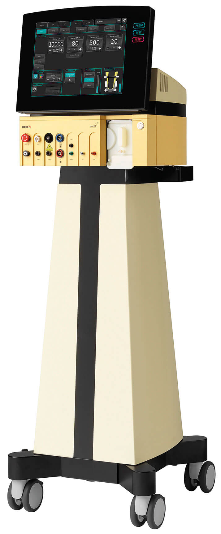
Produced by: Oertli Instrument AGDetail: The SPEEP Mode is a form of peristaltic pump in which the characteristics are reversed – vacuum is controlled by the surgeon, whereas flow is maintained within preset limits. It works in accordance with Poiseuille’s Law, which governs flow rate in a tube. According to the law, vacuum (as controlled by the surgeon) increases flow rate (within preset limits) in a linear fashion. SPEEP mode provides controllable holding force (vacuum), high efficacy, and controlled flow. The advantages of SPEEP mode in combined surgery include controlled lifting of epi-nucleus, aspiration of the cortex (especially when zonules are weak), detaching the posterior hyaloid, and working precisely in the periphery, with or without a detached retina. Impact: SPEEP offers a controllable holding force without flow, and therefore no traction. In combination with the continuous flow-cutter, SPEEP mode is the ideal feature needed for vitreous shaving. Possible new applications could include membrane dissection, aspiration of membranes, aspiration of the subluxated lens particles, and aspiration of subluxated IOLs. One judge said:
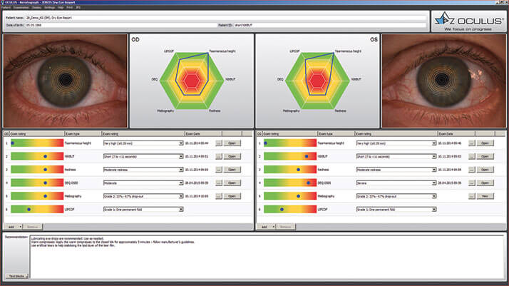
A tool for quickly identifying the cause of dry eye
Produced by: OCULUS Optikgeräte GmbH and JENVIS Research InstituteDetail: Dry eye disease is defined as a multifactorial disease which can cause reduced visual function, optical disturbances and discomfort. But its multifactorial nature means a successful diagnosis may need a combination of tests. The JENVIS Dry Eye Report (JDER) lets users gather all their findings in one overview: the combination of the Dry Eye Questionnaire (DEQ), results of a slit lamp examination, and the noninvasive measurements of the OCULUS Keratograph 5M complete the comprehensive dry eye analysis. The therapy – as well as the treatment plan – can be defined as text blocks so that they can be used and adapted easily for further applications. Impact: Millions of people suffer from dry eye worldwide, and the JDER takes this into account and presents a tool for easy dry eye diagnosis. Providing a way to neatly arrange all findings including values, images, and videos enables easy comparison and integration of findings, the system combines screening and consultancy. The patient can then receive an easy-to-grasp print-out. One judge said:
WIOL-CF: Bioanalogic IOL
A bioanalogic polyfocal IOL for correction of cataract and presbyopia Produced by: Medicem (www.medicem.com) Detail: Building on the heritage of Otto Wichterle, the inventor of hydrogel contact lenses, Medicem has leveraged over 20 years of scientific and clinical research into its proprietary WIGEL hydrogel material that has been specially developed for intraocular applications, creating the WIOL-CF, the first bioanalogic polyfocal IOL for correction of cataract and presbyopia. By mimicking the natural crystalline lens in material, size and design, WIOL-CF is designed to deliver visual quality at all distances. Smooth hyperbolic aspheric optics, with no multifocal refractive or diffractive zones, in combination with biocompatible material allows the patient to perceive a natural transition of vision, while maintaining contrast sensitivity and long-term functionality.
Impact: The market for presbyopia correcting (PC) IOLs has grown dramatically, more than doubling between 2008 and 2012, but PC-IOLs still only represent around 3 percent of total IOLs implanted globally. Many surgeons believe this is due to performance limitations and negative trade-offs such as optical phenomena and low contrast sensitivity, restricting the candidate patients for PC-IOL implantation to only very highly motivated patients who wish to be spectacle-free. Bioanalogic WIOL-CF offers an appealing solution to these problems and holds the potential to substantially grow the PC-IOL market. Judges’ comments: “A new concept of polyfocal IOL to create spectacle independence.”
Orion
Device-independent OCT image analysis software Produced by: Voxeleron (www.voxeleron.com)Detail: OCT is the standard of care in ocular disease management, but is underutilized as current software provided by the OCT manufacturers support measurements of three (of several) retinal layers at most. The retina is an extension of the central nervous system and its deeper, neuronal layers have been shown to help gauge not only the health of the eye, but also to offer direct correlates to brain structure and health. These layers are more challenging to segment, and cannot be measured with existing software. Orion addresses this need with device-independent segmentation of seven retinal layers, including the inner and outer nuclear layers, and has been validated by two independent studies. It also provides automation, speed, and intuitive interaction for an optimized workflow. Impact: Well established in ocular imaging, OCT is now poised to become an important tool in the fight against neurodegenerative diseases including ALS and Alzheimer’s. OCT is likely to become a ubiquitous, front line disease screening and management tool impacting millions of people, but only once analysis software can support it. The technology within Orion is an important step in this direction, that should help accelerate the pace of discovery in ophthalmology and neuroscience, and empower clinical researchers to study the relationship between the neuronal layers of the retina and a wide variety of neuropathies. Judges’ comments: “Good alternative in hospitals with different OCT manufacturers to have same software for evaluation.”
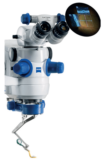
OPMI LUMERA 700 and RESCAN 700
Providing ophthalmic surgeons ZEISS HD-OCT images of the eye without interrupting surgeryThe RESCAN 700 has a broad range of applications in anterior and posterior segment surgery.ZEISS HD-OCT images add a real-time third dimension to the visualization capabilities directly in the eyepiece of the OPMI LUMERA 700 surgical microscope. It provides surgeons with unprecedented views below the surface of the surgical field, enabling them to see more – even transparent structures – and helping them back up their decisions and improve their surgical technique, without compromising surgical workflow. By merging two gold standards into one system, ZEISS has created a first – a surgical microscope with integrated intra-operative OCT: the OPMI LUMERA 700 and RESCAN 700. Impact: Today’s surgeons may have difficulty seeing certain anatomic details during surgery. With this new visualization tool, ophthalmologists can overcome these limits. Surgeons now can see even transparent ocular structures during surgery, monitor progress during a procedure, and verify clinical results in the OR. OCT scans can also be stored and recalled for later review and “fly through” via CALLISTO eye from ZEISS. Simply put, the new device enables better decision-making during surgery. Judges’ comments: “Enables direct control of both anterior and posterior segment procedures.”
OCULUS BIOM ready
The world’s first single-use wide angle viewing system Produced by: Oculus Surgical (www.oculussurgical.com)Detail: Provides the perfect view for non-contact wide angle observation for retinal surgeons without using a contact lens. Easily connected to the microscope; while the surgeon is observing the vitreous and the fundus, the BIOM ready is aligned coaxially with the operating microscope… but during extraocular surgery phases, it’s swung out of the observation beam. It incorporates the new BIOM HD Disposable Lens for unparalleled visual clarity and provides excellent depth of field for better stereopsis. The OCULUS BIOM ready comes pre-assembled with the BIOM HD Disposable Lens in a sterile blister pack. Impact: Provides the optimal balance between efficiency and high optical quality, with outstanding resolution in the periphery, often reducing indentation during laser. The depth of field is increased over other wide angle systems, allowing the surgeon to perform macula work without the need for a contact lens, saving time and money. Sterilization “down time” is decreased – increasing OR efficiency – and as it’s a single-use device, it reduces the risk of cross-contamination. Judges’ comments: “Amazing wide field view in a disposable lens.” “Improves convenience.”
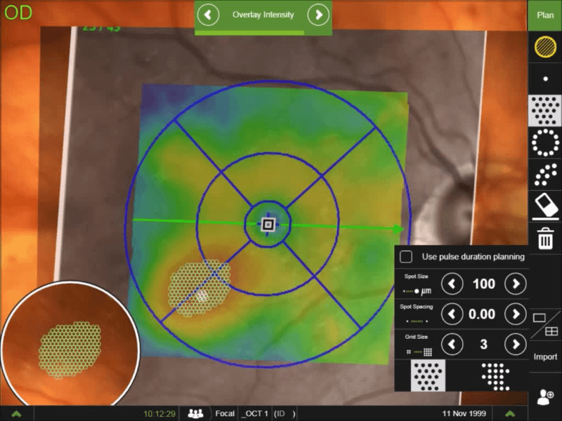
Navilas 577+
Tissue-friendly, standardizable, navigated microsecond pulsing therapy for retinal disease Produced by: OD-OS (www.od-os.com)Detail: Laser energy is split up into a pulse train of low-energy pulses that stimulate the retina, but do not heat the tissue to a coagulation threshold – and as a consequence, retinal function loss and scarring can be avoided. Navilas is the only navigated microsecond pulsing therapy (NMPT) system that allows navigated application of this advanced subthreshold laser technique; the treatment area can be precisely delineated based on imported OCT thickness maps, and the aiming beam is prepositioned – compensating for eye movement and allowing complete coverage without undefined overlap. The treatment is documented in real time, providing visual feedback about treated areas and degree of completion. An initial case series at LMU Munich performed by Marcus Kernt showed no tissue damage or retinal function loss (as expected) with this method. Contact lens-free application and comfortable infrared illumination make this a patient-friendly therapy and set Navilas 577+ apart from slit lamp-based lasers. Impact: Retinal laser is making a comeback in diabetic eye disease because of the chronic use and expense associated with anti-VEGF therapy. Initial studies indicate that laser treatment can add durability to anti-VEGF gains and reduce patient burden. The technique further refines retinal laser therapy: inter-operator variability is minimized by OCT-based planning with real-time documentation, and retinal tissue function is preserved by microsecond pulsing. NMPT has the potential to become the standard adjunct to anti-VEGF in diabetic eye disease. Judges’ comments: “Great laser to improve patients’ outcomes.”