Conexiant
Login
  • Corneal Physician
  • Glaucoma Physician
  • New Retinal Physician
  • Ophthalmology Management
  • Ophthalmic Professional
  • Presbyopia Physician
  • Retinal Physician
The Ophthalmologist
  • Explore

    Explore

    • Latest
    • Insights
    • Case Studies
    • Opinion & Personal Narratives
    • Research & Innovations
    • Product Profiles

    Featured Topics

    • Anterior Segment
    • Glaucoma
    • Retina

    Issues

    • Latest Issue
    • Archive
  • Subspecialties
    • Cataract
    • Cornea
    • Glaucoma
    • Neuro-ophthalmology
    • Oculoplastics
    • Pediatric
    • Retina
  • Business

    Business & Profession

    • Professional Development
    • Business and Entrepreneurship
    • Practice Management
    • Health Economics & Policy
  • Training & Education

    Career Development

    • Professional Development
    • Career Pathways

    Events

    • Webinars
    • Live Events
  • Events
    • Live Events
    • Webinars
  • Community

    People & Profiles

    • Power List
    • Voices in the Community
    • Authors & Contributors
  • Multimedia
    • Video
    • Podcasts
Subscribe
Subscribe

False

Advertisement
The Ophthalmologist / Issues / 2013 / Dec / Smart(phone) Ophthalmoscopy
Business and Entrepreneurship

Smart(phone) Ophthalmoscopy

An iPhone, an app, and a 20 D lens, along with a bit of practice, will get you fundus photographs in a pinch

By Mark Hillen 12/2/2013 1 min read

Share

0313-203 fig.1Figure 1. a. The Welch Allyn iExaminer device. Figures 1b-d. Handheld 20D lens imaging of: b. Familial exudative vitreoretinopathy imaged during examination under anesthesia; c. Large choroidal nevus imaged in an Emergency Department setting; d. Partially treated retinoblastoma imaged during examination under anesthesia.

If you think that taking images of the retina requires an ophthalmology clinic decked out in expensive instrumentation – a condensing lens and coaxial light source, a dedicated image acquisition device, all linked to a computer – think again. The rise of smartphones that can shoot high-definition video, take even higher-resolution still images and provide a constant light source from a LED flash, means that everything but the specialized optics can be accomplished with a high-end iPhone or equivalent.

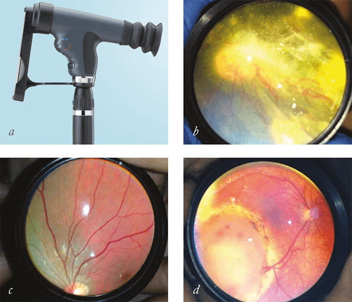
These recent capabilities haven’t escaped the attention of medical diagnostic equipment manufacturers. One, Welch Allyn, lets you pair their €540 PanOptic Ophthalmoscope with an iPhone 4 or 4S with a professional-looking €65 adaptor and its associated free app (Figure 1a). Image acquisition, storage, and distribution are all performed on the smartphone – increasing convenience and cutting costs (the total purchase price excluding the iPhone is only €605). Now, three Boston-based ophthalmologists have lowered the bar even further. Luis Haddock, David Kim and Shizo Mukai of the Massachusetts Eye and Ear Infirmary have come up with a solution that is cheaper and more portable (1). It involves holding a €20, 20 D lens in front of the eye, and using the iPhone to video the images of the lens (Figure 1b–d).

When this has been tried before, results were unsatisfactory, primarily because the in-built Apple iPhone Camera app was poor. Apple’s app did not to allow users to adjust focus or contrast whilst filming, leading to blurry images that were full of glare. Luis, David and Kim’s solution is simple: buy a better camera app. They chose FiLMiC Pro, which currently retails for €2.99 on the Apple iPhone App Store. It enables focus and exposure adjustments during filming. The results are great – both with and without Koeppe lens use, and the group have been able to capture excellent, high-quality fundus images in children under anesthesia and in awake adults. “Our technique provides a simpler and higher-quality method to more consistently produce excellent images of a patient’s fundus,” said Shizuo Mukai, explaining that “this technique has been extremely helpful for us in the Emergency Department setting, in in-patient consultations, and during examinations under anesthesia, as it provides a cheaper and portable option for high-quality fundus-image acquisition for documentation and consultation”. Looking to justify the purchase of the latest smartphone? Upgrading your iPhone is likely to improve the camera in the device – and with it, the quality of your ophthalmoscopy

References

  1.  LJ Haddock, DY Kim, S Mukai, “Simple, Inexpensive Technique for High-Quality Smartphone Fundus Photography in Human and Animal Eyes”, J. Ophthalmol, Article ID 518479 (2013)

About the Author(s)

Mark Hillen

I spent seven years as a medical writer, writing primary and review manuscripts, congress presentations and marketing materials for numerous – and mostly German – pharmaceutical companies. Prior to my adventures in medical communications, I was a Wellcome Trust PhD student at the University of Edinburgh.

More Articles by Mark Hillen

Related Content

Newsletters

Receive the latest Ophthalmology news, personalities, education, and career development – weekly to your inbox.

Newsletter Signup Image

False

Advertisement

False

Advertisement

Explore More in Ophthalmology

Dive deeper into the world of Ophthalmology. Explore the latest articles, case studies, expert insights, and groundbreaking research.

False

Advertisement
The Ophthalmologist
Subscribe

About

  • About Us
  • Work at Conexiant Europe
  • Terms and Conditions
  • Privacy Policy
  • Advertise With Us
  • Contact Us

Copyright © 2025 Texere Publishing Limited (trading as Conexiant), with registered number 08113419 whose registered office is at Booths No. 1, Booths Park, Chelford Road, Knutsford, England, WA16 8GS.

Disclaimer

The Ophthalmologist website is intended solely for the eyes of healthcare professionals. Please confirm below: