
Kai Jin (Credit: Headshot supplied by author).
The retina stands as a quintessential component of the central nervous system (CNS) (1). Nobel Prize-winner and father of modern neuroscience, Santiago Ramón y Cajal, was – very rightly – fascinated with the retina, and his pioneering work and artistic, hand-drawn representations in the late nineteenth century highlighted its complexity and connectivity within neural structures. Cajal’s devotion to studying the retina underscored its significance as a fundamental part of sensory processing and neural connectivity, and his teachings continue to emphasize the clinical relevance of the visual pathway, reaffirming the retina’s vital role in the intricate neural network of the CNS (2).
We should also mention here that another major contributor to this area, Polish ophthalmologist Ksawery Gałęzowski (1832-1907), who in 1866 published a textbook on the value of examining the optic disc to diagnose CNS disorders (3), coining the term “cerebroscopy” in the process. Because of his collaborations with famed neurologist Jean-Martin Charcot at the Salpetriere in Paris, Gałęzowski gained a great deal of experience with neurological diseases. An immigrant to France, in 1865 he was given the title of Doctor of Medicine with his doctoral dissertation, “On Pathologic Changes of the Optic Nerve and Cerebral Diseases from which They Originate.”
In the realm of neurology, precise and non-invasive imaging techniques are paramount for understanding the pathophysiology of diseases and devising effective therapeutic strategies. Optical coherence tomography (OCT) has garnered increasing attention as a high-resolution imaging technique within the field of neuroscience (4); for the study of CNS diseases, the modality exhibits unique potential. It also holds promise in neurodegenerative conditions, such as multiple sclerosis and Alzheimer’s disease (AD) – see Figure 1 (5). Through OCT, observations of neuronal damage, brain atrophy, and alterations in nerve fibers are possible – all of which are often associated with the progression and development of these diseases. Moreover, OCT’s capability for dynamic imaging enables the tracking of pathological changes in neurodegenerative diseases, potentially serving as a robust tool for evaluating treatment efficacy and disease progression (see Table 1).

Andrzej Grzybowski (Credit: Headshot supplied by author).
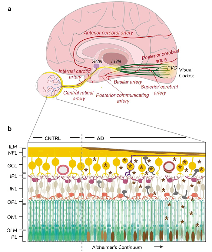
Figure 1. Retinal projections into the brain and AD pathological progression in retinal tissue.
Table 1. OCT has been used across various CNS disorders, providing insights into retinal changes and structural alterations associated with these conditions.
Within ophthalmology, eye-based AI algorithms are emerging as a revolutionary tool in diagnosing and monitoring CNS disorders (5). Leveraging sophisticated technology and image analysis, these algorithms use data from eye scans, particularly from techniques like OCT, to detect subtle changes in the retina. By detecting anomalies in retinal structure, vasculature, or cellular composition, they offer early indicators for various CNS disorders, including neurodegenerative diseases like AD and Parkinson’s disease (7,8). The integration of eye-based AI algorithms into clinical practice holds promise for early disease detection, tracking disease progression and evaluating treatment responses. This progress represents a fusion of the retina’s importance with cutting-edge technology, ushering in a new era where the eye serves as a diagnostic gateway to unravel complexities within the CNS, potentially revolutionizing how we understand and manage neurological disorders.
In conclusion, OCT stands as a fast, non-invasive, and high-resolution imaging technique with broad applications in the study and clinical diagnosis of neurological diseases. By combining various imaging modalities, a more comprehensive and accurate characterization of CNS diseases can be obtained, thereby supporting early diagnosis, monitoring, and treatment of these conditions (9). Looking ahead, with ongoing technological advancements and further clinical research, OCT is poised to become an indispensable imaging tool in the field of neuroscience.
More on this topic can be found in our book, OCT and Imaging in Central Nervous System Diseases. The Eye as a Window to the Brain (10).
References
- P Frith, AR Mehta, “The retina as a window into the brain,” The Lancet Neurology, 20, 892 (2021). PMID: 34687632.
- SRY Cajal, “Advice for a young investigator,” MIT Press: 2004.
- X Gałęzowski, “Etude ophtalmoscopique sur les altérations du nerf optique et les maladies cérébrales dont elles dépendent,” Paris: Librairie de L. Leclerc: 1866.
- HM Kersten et al., “Ophthalmic manifestations of inherited neurodegenerative disorders,” Nature Reviews Neurology, 10, 349 (2014). PMID: 24840976.
- J Wolf et al., “Liquid-biopsy proteomics combined with AI identifies cellular drivers of eye aging and disease in vivo,” Cell, 186, 4868 (2023). PMID: 37863056.
- V Parisi et al., “Correlation between morphological and functional retinal impairment in multiple sclerosis patients,” Invest Ophthalmol Vis Sci, 40, 2520 (1999). PMID: 10509645.
- BA Nichol, AC Hurlbert, “Predicting attitudes towards screening for neurodegenerative diseases using OCT and artificial intelligence: Findings from a literature review,” J Public Health Res, 11, 4 (2022). PMID: 36310821.
- X Wang et al., “Machine learning based on Optical Coherence Tomography images as a diagnostic tool for Alzheimer’s disease,” CNS Neurosci Ther, 28, 2206 (2022). PMID: 36089740.
- D Li et al., “Deep learning in optical coherence tomography: Where are the gaps?” Clinical & experimental ophthalmology, 51, 853 (2023). PMID: 37245525.
- A Grzybowski, P Barboni (eds.) OCT and Imaging in Central Nervous System Diseases. The Eye as a Window to the Brain, eds, Springer Cham: 2020.
