Sponsored by
Convergent Evolution: A Lesson From the Eye of Marsupials
Many ophthalmologists probably have a human-centric view of ocular anatomy. Indeed many likely think that all mammals have a retinal blood supply like ourselves - but of course this is not true. Many mammals, such as grazing animals, have fairly simple and thus thin retinae that do not have direct intraretinal vessels and rely on the choroid for all their nutrition. Now one would imagine that in species with retinal vessels, they would be arranged in approximately the same pattern in all mammals.Well it appears that when mammals split roughly 130 million years ago into eutherian (placental) mammals and marsupials, things got interesting... Marsupials also have groups with simple retinae (including kangaroos, wombats, koalas and possums) and some with complex retinal functions (numbats, opossums and the Tasmanian devil) that evolved a retinal blood supply. However, as can be seen in this example of a South American opossum (Monodelphis domestica) (a) when one looks at their retina with a fundus camera it looks (at first) like a mouse retina (b). However if one looks closely at the fluorescein angiogram (arrow in (c)) one can see there are in fact two vessels and not one. Histology (d) shows that these pairs of vessels consist of an artery on the vitread aspect of the corresponding vein. This pattern can be seen in a flat mount of a retina that has been perfused with fluorescent dye (e). Here you can see the pairs of vessels and how they terminate in numerous hairpin loops. This pattern is completely different from placental mammals where capillaries form anastomotic beds and arteries/arterioles and veins/venules lie apart from one another except where they enter the eye at the optic nerve head. So whilst the marsupials with complex retinal functional needs have solved the issue of providing a blood supply, they have used a different, double vascular pattern that is only seen in marsupial retinae and brains. There appears no obvious functional advantage to this pattern and it seems to be a classic example of convergent evolution. Paul McMenamin, Director of the Centre for Human Anatomy Education, Monash University, Melbourne, Australia.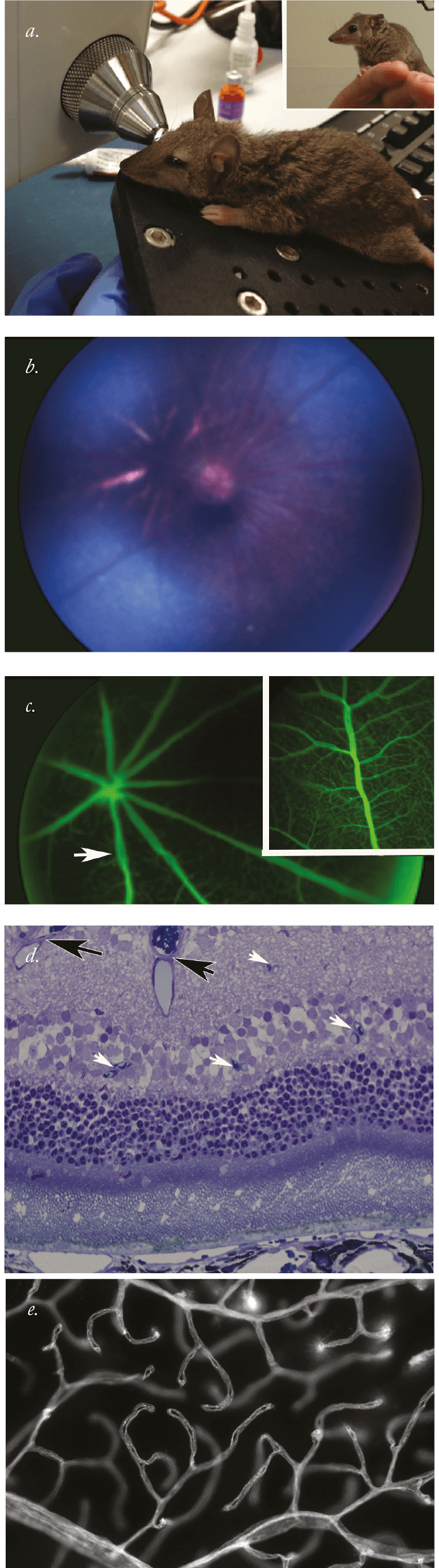
The Light of the Eye
This image was created in response to the Lawrence Ferlinghetti poem “Instructions to painters and poets”: … and don’t forget to paint all those who lived their lives as bearers of light… paint the light of their eyes… Gianpiero Actis, Eye surgeon and award-winning artist, Torino, Italy.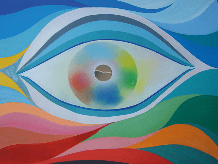
Eyelash Mites
Colored scanning electron micrograph of an eyelash hair growing from the surface of human skin; the tails of eyelash mites are seen protruding from the base. The eyelash mite or Demodex folliculorum is a parasite found in the follicles of the human face, mainly in the nose, cheeks and most especially the eyelash area. Steve Gschmeissner, scientific photographer, Bedford, UK.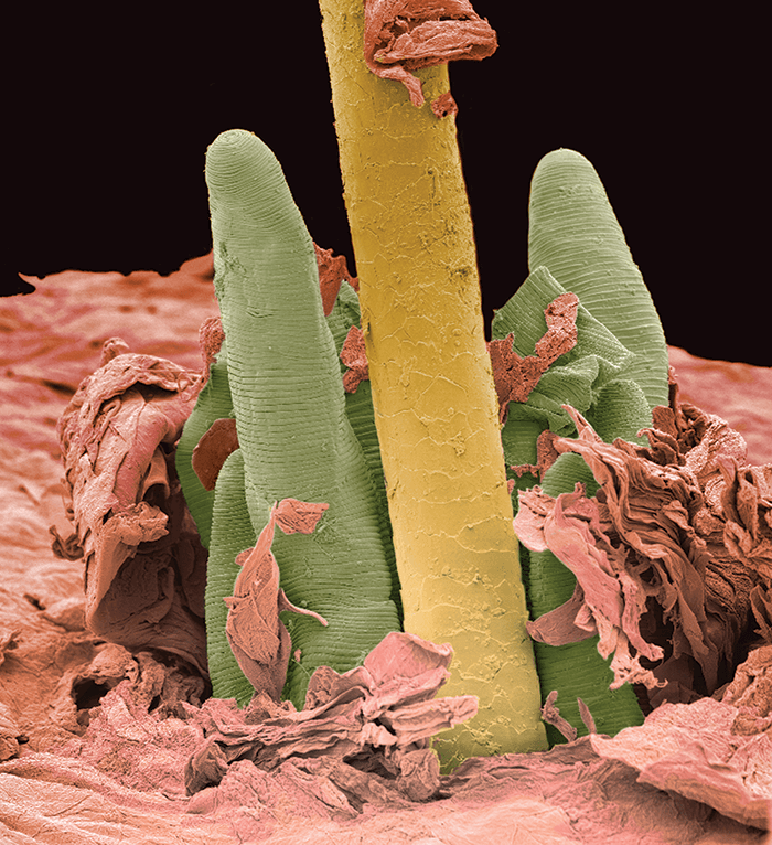
Coloboma
A patient with typical peripheral retinal coloboma. Kelly Aileen Oldstein, Certified Ophthalmic Photographer at Chester, County Eye Care and Owner, Kelly Aileen, Photography, West Chester, PA, USA.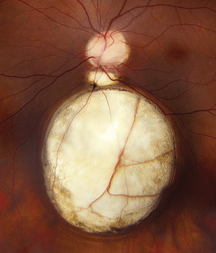
Posterized Posterior Segment
Noninvasive visualization of the optic nerve head and peripapillary vascular network using optical coherence tomography angiography (OCTA) (Optovue RTVue XR Avanti, Optovue, Inc., Fremont, CA). A) Fundus photograph of the left eye in a healthy control. B) 10 frames averaged OCTA color overlay with C) superficial vessels including radial peripapillary capillaries in red and D) deeper vessels including major arteries and veins in cyan. Shelley Mo, Toco YP Chui and Richard B Rosen, New York Eye and Ear Infirmary of Mount Sinai, New York City, NY, USA.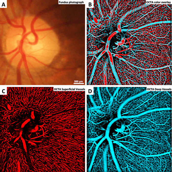
Bird’s Eye View
The normal eye of a red-tailed hawk. The Comparative Ocular Pathology Laboratory of Wisconsin (COPLOW), USA. Further images can be found on the COPLOW Facebook page: bit.ly/COPLOW
A Dog’s Life
A dog eye with early-life trauma, which is believed to have happened prior to the eyelids opening at 12 days of life. Image courtesy of COPLOW.
Helianthus
A pupil surrounded by a yellow-pigmented rich region of the iris, reminiscent of a sunflower. Kelly Aileen Oldstein, Certified Ophthalmic Photographer at Chester County Eye Care and Owner, Kelly Aileen Photography, West Chester, PA, USA.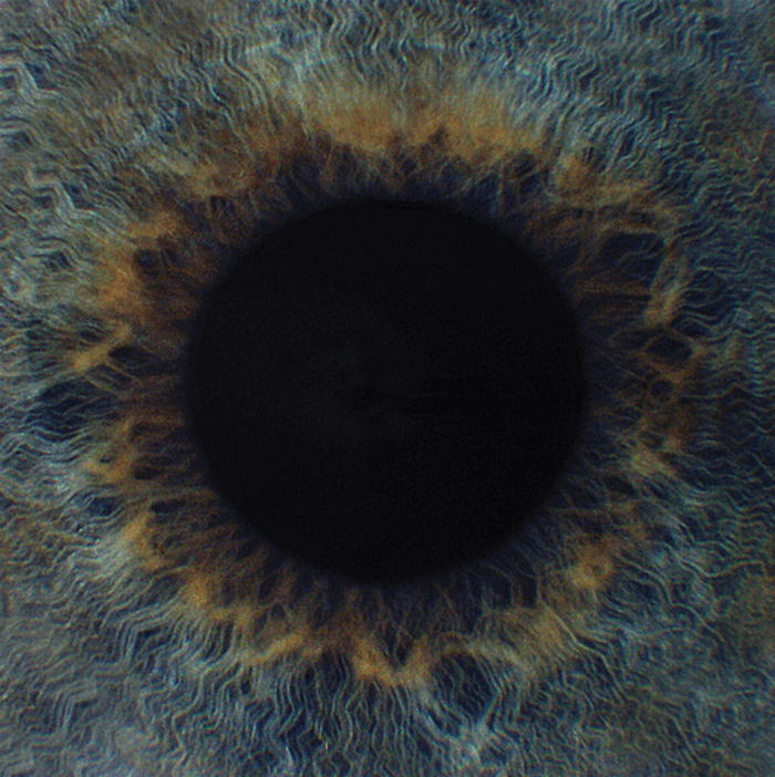
Deadeye
Experimental setup for puncture testing of full human cadaveric globe specimens. From: Andrew Rau, Scott Lovald, Steven Nissman, John McNulty, Jorge Ochoa, and Michael Baldwinson. “The mechanics of corneal deformation and rupture for penetrating injury in the human eye”, presented at ARVO 2016.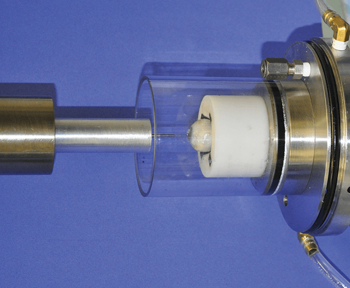
Web of Intrigue
Segmentation of superficial vascular plexus in a neonatal mouse retina. Sabine Uhles, Franco Revelant, Sabine Grüner, Guido Hartmann and Fethallah Benmansour, Ocular Discovery and Biomarkers, Roche, Basel, Switzerland.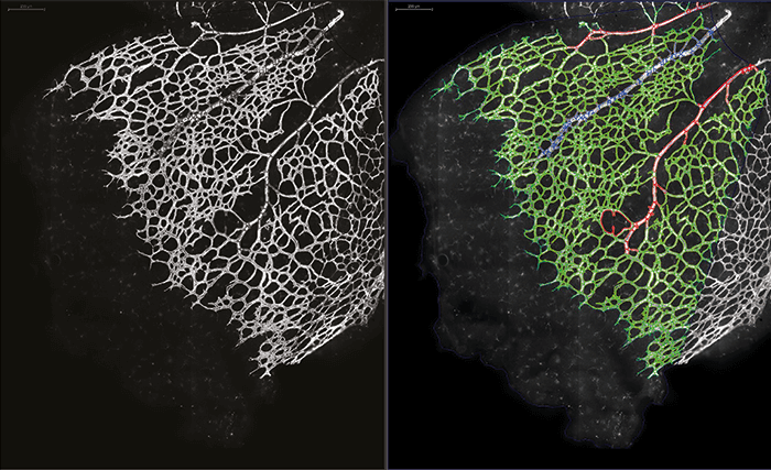
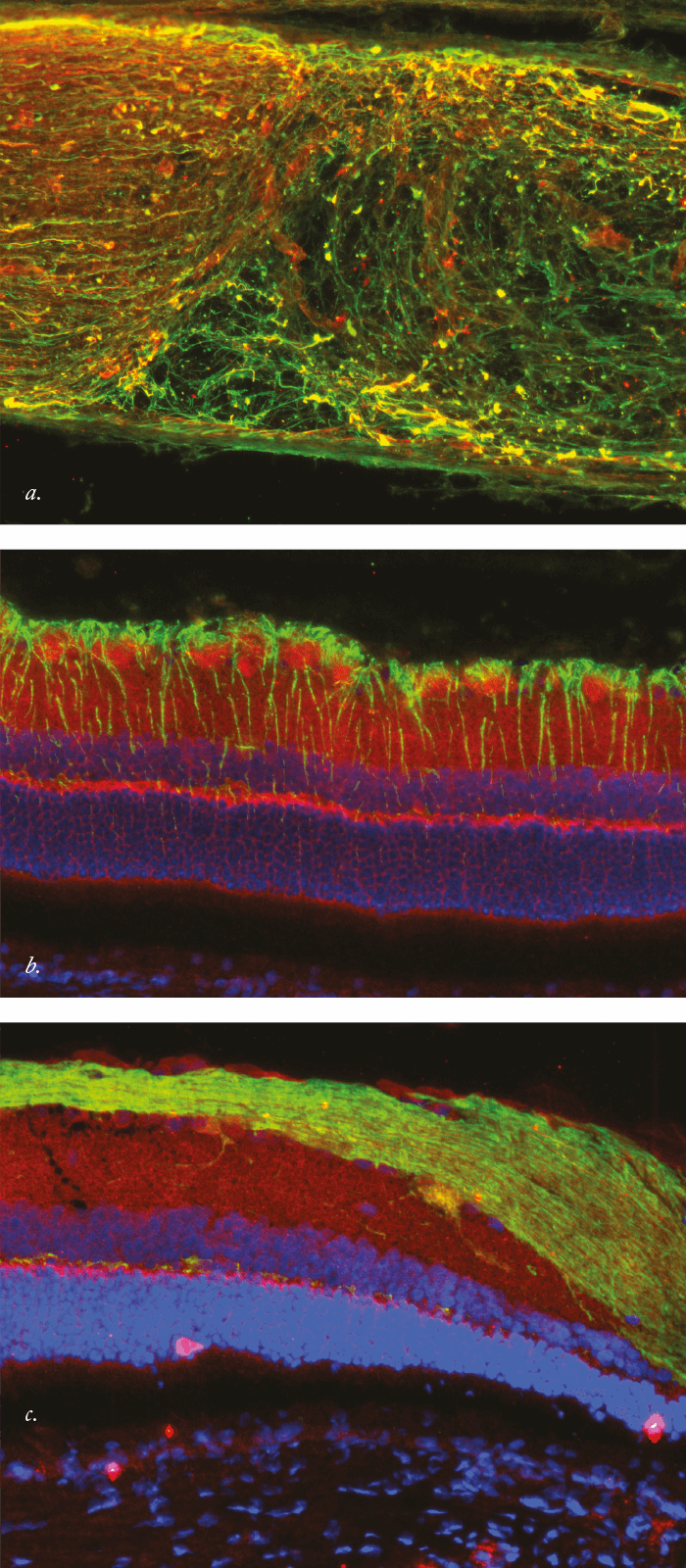
Illuminating Immunohistochemistry
Confocal microscopy imaging of rat retinal sections. a. Double staining of a crushed optic nerve. Nerve fibers are stained in green, and the repulsive guidance molecule A is stained in red. The picture is taken 10 days after optic nerve crush directly at the crush site. b. Triple staining of rat retina after optic nerve crush with cell nuclei in blue, GFAP-positive cells in green and repulsive guidance molecule A staining in red. c. Triple staining of rat retina after optic nerve crush with cell nuclei in blue, nerve fibers and RGCs in green and repulsive guidance molecule A staining in red. Sven Schnichels, Laboratory Head at the University Eye Hospital, Tübingen, Germany.