Sponsored by
X Marks the Spot
This female patient presented with X-linked retinitis pigmentosa with symptoms including decreased night vision and mild glare. Female carriers often display a reduced electroretinogram signal, but tend not to develop other complications. Kelly Aileen Oldstein, Certified Ophthalmic Photographer at Chester County Eye Care and Owner, Kelly Aileen Photography, West Chester, PA, USA.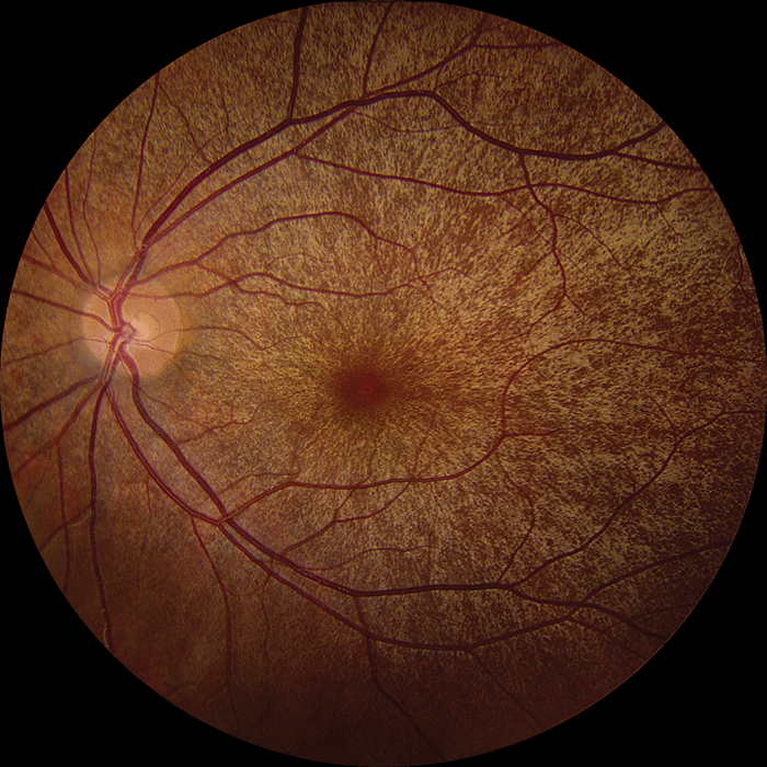
Tesla Coil
This wide-angle fluorescein angiogram shows severe proliferative diabetic retinopathy. Kelly Aileen Oldstein, Certified Ophthalmic Photographer at Chester County Eye Care and Owner, Kelly Aileen Photography, West Chester, PA, USA.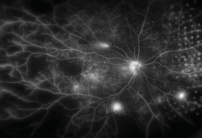
486 nm + 815 nm = ...
The infrared image shows an odd splotching pattern, but a quick glance with fundus autofluorescence displayed a completely different view of the pathology. Houston Sharpe III, Ophthalmic Imaging Specialist, UNC Kittner Eye Center, Chapel Hill, NC, USA.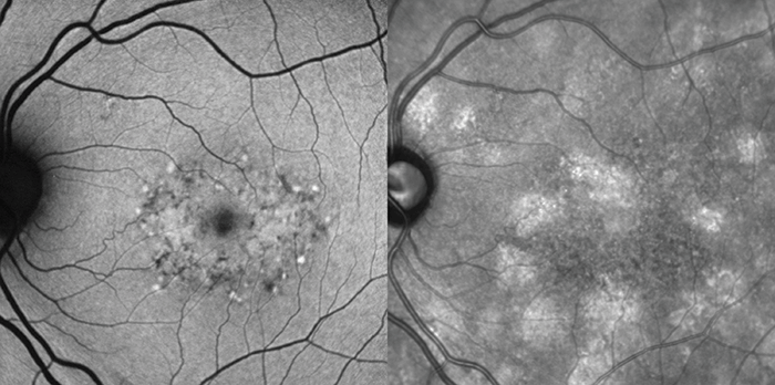
Forbidden Planet
Choroideremia is a rare disease with a strong genetic component. Progressive vision loss results from degeneration of the retinal pigment epithelium, and the choriocapillaris is clearly demonstrated in this widefield fluorescein angiogram. Joseph Territo, Ophthalmic Photographer, Retina Associates of Western New York, Rochester, NY, USA.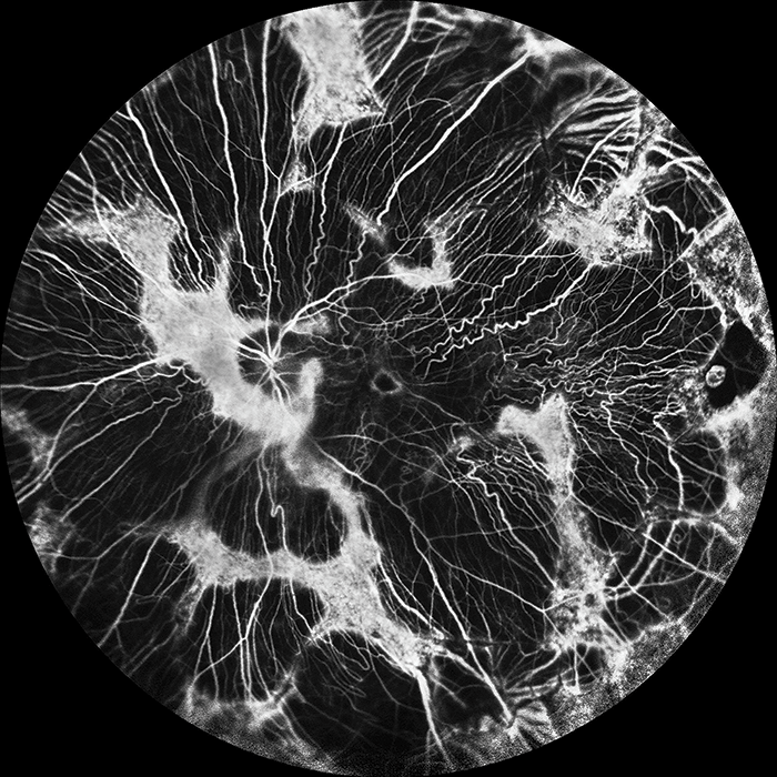
Retinal Nebula
Severe proliferative diabetic retinopathy with laser treatment. Karen Gasperian, Ophthalmic Photographer at Retina-Vitreous Associates Medical Group, Los Angeles, California, USA.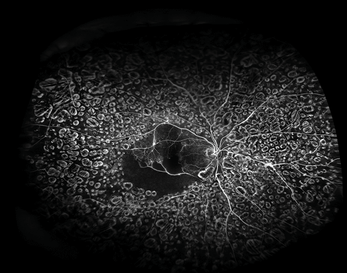
Proximity Fuze
Color image of a patient’s fundus displaying torpedo maculopathy. Karen Gasperian, Ophthalmic Photographer at Retina-Vitreous Associates Medical Group, Los Angeles, CA, USA.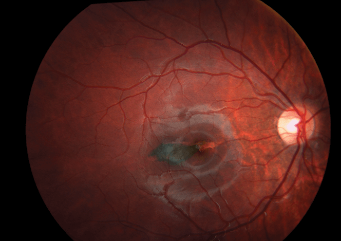
Moon Through Trees
A strikingly high-contrast angiograph makes for an incredibly artistic image of the eye. Kelly Aileen Oldstein, Certified Ophthalmic Photographer at Chester, County Eye Care and Owner, Kelly Aileen Photography, West Chester, PA, USA.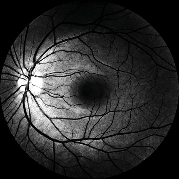
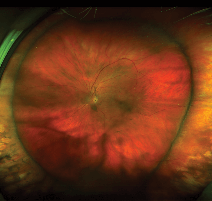
Scleral Buckling Surgery
Ultrawide-field color image of the left fundus of a 58-year old female patient who underwent a scleral buckling surgery to correct a macula-off rhegmatogenous retinal detachment. The encircling band and 360° of moderate scleral indentation are visible. A. Osman Saatci, Dokuz Eylul University, İzmir, Turkey.